Embryonic Development Of A Frog
Studying embryonic vertebrate development in the frog is useful because the frog possesses all of the basic characteristics of nonamphibious vertebrates. Because the frog embryo develops externally, this process can be easily observed. The egg is large enough to be visible to the naked eye and develops quickly, making the study of the frog's embryonic development one that can be carried out over a short period, typically between 12 to 16 weeks.
The Egg and Fertilization
The Egg and Fertilization
Frogs lay many eggs in a mass or spawn, which serves to protect most of the eggs from predators. The male frog fertilizes the eggs as the female lays them in water. That is, the eggs are fertilized outside the female's body. Each frog egg is a single cell but is an unusually large one that is visible to the human eye. As the fertilized egg, or zygote, goes through its life cycle, the resultant complete tadpole will contain many millions of cells but will be essentially the same size and weight as the progenitor egg cell. In effect, the single cell develops into a multicellular tadpole.
The Cleavage and Blastula Stage
The Cleavage and Blastula Stage
Cleavage is the process of cell division in the early embryo. The frog zygote undergoes rapid cell division without experiencing overall growth, resulting in a cluster of cells the same volume and mass as the original zygote. The different cells derived from cleavage are called blastomeres and form a compact mass called the morula. The blastula stage occurs when a hollow ball of cells forms around a cavity filled with fluid.
The Gastrulation Process
The Gastrulation Process
The typical blastula is merely a ball of cells. The next stage in frog embryonic development represents a big leap forward: It comprises the formation of the planned shape and structure of the animal, known as the body plan. The cells in the blastula rearrange themselves to form three layers of cells in a process called gastrulation. During gastrulation, the blastula forms these three layers of cells, called germ layers, which will differentiate into different organ systems.
Cell Differentiation
Cell Differentiation
As cells begin to differentiate, they are said to be "fated," meaning each has particular purposes associated with it. The three germ layers are the endoderm, the ectoderm and the mesoderm. The ectoderm gives rise to the nervous system and the skin; the mesoderm forms muscle cells, internal organs and connective tissue; and the endoderm ultimately forms the types of cells found in the digestive system, lungs and many internal organs.
The Tadpole's Growth and the New Frog
The Tadpole's Growth and the New Frog
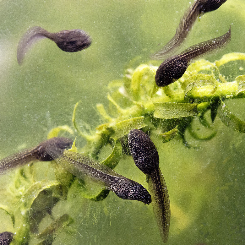
In time, the egg hatches, and the result is an independent living creature called a tadpole — the aquatic larval stage of a frog — with gills, mouth and tail. Over a period of one to three months, the tadpole will begin to change to the amphibious frog, with lungs replacing gills, a gradual shortening of the tail and the appearance of legs. After about 12 weeks, its tail is nearly gone and it is able to leave the water. By 16 weeks or so, the new frog is able to begin the reproductive process.
Cite This Article
MLA
Beacom, Betsy. "Embryonic Development Of A Frog" sciencing.com, https://www.sciencing.com/embryonic-development-frog-6711996/. 13 March 2018.
APA
Beacom, Betsy. (2018, March 13). Embryonic Development Of A Frog. sciencing.com. Retrieved from https://www.sciencing.com/embryonic-development-frog-6711996/
Chicago
Beacom, Betsy. Embryonic Development Of A Frog last modified March 24, 2022. https://www.sciencing.com/embryonic-development-frog-6711996/