How Is A Sample Prepared For Viewing Under A Microscope?
A formerly invisible realm was revealed in the early 1600s with the construction of the first compound microscopes led to major revisions in scientific understanding. Basic compound microscopes are now standard equipment in medicine and natural sciences. Transmitted visible light shines through thin preparations for magnification. Transmission and scanning electron microscopes developed from 1931 onwards. They don't use optical light, but beams of electrons and magnetic fields to view specimens. Mainly for institutional research, specimen preparation requires complex, expensive equipment.
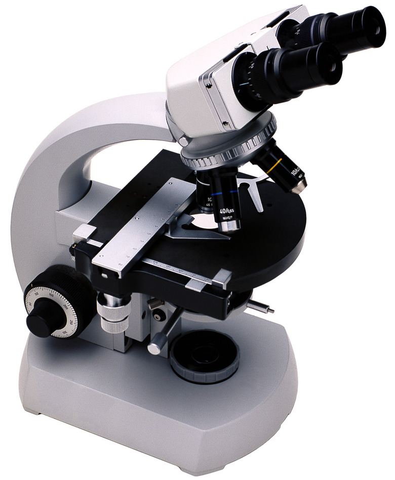
Understanding Compound Microscopes
Understanding Compound Microscopes
Several specialized types of compound microscopes exist, but bright field microscopes are most common. Samples for them should be only several microns, which is a millionth of a meter, thick. Thicker specimens don't let enough light through and don't allow precise focusing. Bright field microscopes have a tube with objective lenses at the bottom, closest to the specimen, and an ocular lens, or eyepiece, at the top. Several objective lenses of different magnifications revolve on a nosepiece, or turret. The stage just below the nosepiece holds the specimen slide, and below that a source of light shines up through a condenser to the specimen. Modern compound microscopes can magnify an object from 1,000 to 2,000 times its original dimensions.
Whole Mounts
Whole Mounts
For small items such as hairs, small insects, insect parts or pollen grains, the sample is placed directly on the center part of a glass or plastic microscope slide with a small amount of mounting medium, usually a synthetic or natural resin product for permanent slides. For temporary slides, such as a drop of pond water containing microorganisms, the water is the mounting medium. Protect samples with a cover slip, a round or square very thin piece of glass or plastic. Some samples need staining with natural or synthetic dyes meant for microscopy to be seen well.
Squashes and Smears
Squashes and Smears
A simple way to prepare a thin sample is to squash or flatten a small piece of tissue under the cover slip. Often used in plant samples to see chromosomes, rapidly growing tissues such as root tips or anthers undergoing cell division are preserved in fixative, then softened and stained to reveal the chromosomes. Gentle pressure from the eraser end of a pencil centered over the cover-slipped sample forces the cells apart into a single layer. In smears, the sample is spread thinly across one slide using another slide as the spreader, and the resultant smear is dried and stained. In medicine, samples of bodily fluids, such as blood, cerebro-spinal fluid or semen are smeared.
Stained Tissue Sections
Stained Tissue Sections
A more complicated sectioning procedure occurs when the structure and organization of a whole small organism or a tissue piece needs study. For most samples, first the tissue is preserved and hardened and the water removed. Then the sample is embedded in a rigid medium such as wax or plastic and sliced into very thin sections only several microns thick using a precision machine called a microtome. The sample is oriented to give cross-sections or longitudinal sections when sliced. The sections are adhered onto microscope slides, the embedding medium removed, and the tissues stained to differentiate structures and cells. Where speed is essential, such as in surgical biopsies for cancer, samples are frozen, sliced with a freezing microtome, stained and examined.
References
- Miami University College of Arts and Science: Founding Fathers of Microscopy
- Leica Biosystems: Pathology Leaders: An Introduction to Specimen Preparation
- Miami University College of Arts and Science: Types of Microscopes
- John Innes Centre: Microscopy: What is Electron Microscopy?: The History of EM
- Rice University: Experimental Biology: Light Microscopy
- Miami University College of Arts and Science: The Compound Light Microscope
- Marietta College: Mitosis in Onion Root Tip Cells
Cite This Article
MLA
Csanyi, Carolyn. "How Is A Sample Prepared For Viewing Under A Microscope?" sciencing.com, https://www.sciencing.com/sample-prepared-viewing-under-microscope-9290/. 24 April 2017.
APA
Csanyi, Carolyn. (2017, April 24). How Is A Sample Prepared For Viewing Under A Microscope?. sciencing.com. Retrieved from https://www.sciencing.com/sample-prepared-viewing-under-microscope-9290/
Chicago
Csanyi, Carolyn. How Is A Sample Prepared For Viewing Under A Microscope? last modified August 30, 2022. https://www.sciencing.com/sample-prepared-viewing-under-microscope-9290/