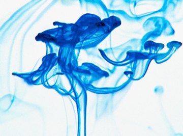
A microbiologist uses chitin staining so she can see fungi clearly under the microscope. Fungi use chitin as structural material in their cell walls so the stain shows the cell wall well. The lactophenol cotton blue stain is the most common stain for fungi. The phenol kills microorganisms and prevents fungal enzymes from breaking down the cell. The cotton blue dye turns the chitin blue. Chitin staining is a relatively straightforward procedure with inexpensive materials. You can either make your own dye or buy commercially prepared stain.
- 0.05 grams cotton blue dye
- 20 g phenol crystals
- 40 milliliters glycerol
- 20 ml lactic acid
- 20 ml distilled water
- 1 piece filter paper
- Weighing scales
- Test tube
- 250 ml beaker
- 1 microscope slide
- 2 to 3 sterile droppers
- 1 cover slip
- 5 ml 70 percent alcohol solution
- 1 sterile loop or 1 bottle commercially prepared lactophenol cotton blue dye
Prepare a solution of lactophenol cotton blue. Weigh out 0.05 grams of cotton blue dye using a sensitive weighing scale. Mix the dye with 20 milliliters of distilled water in a test tube and leave it overnight.
Put on gloves before finishing the stain preparation the next day. Place 20 ml of lactic acid into a beaker and mix it with 20 g of phenol crystals. Stir until the mixture is dissolved. Add in 40 ml of glycerol and mix. Then strain the cotton blue solution through a piece of filter paper into the beaker and mix thoroughly.
Alternatively, use a commercially prepared source of the stain. Store the stain at room temperature.
Label a microscope slide with the sample identification name or number. Use one sterile dropper to put a single drop of a water sample onto the center of the microscope slide. For a dry sample, place a single drop of 70 percent alcohol solution onto the slide, and then use a sterile loop to mix the dry sample into the alcohol.
Use a clean dropper to transfer two drops of the lactophenol cotton blue stain onto the center of the slide. If you are using 70 percent alcohol on the slide, do this before the alcohol evaporates off the slide.
Touch one edge of the microscope cover slip to one edge of the wet sample preparation. Let the slip fall gently onto the sample preparation, ensuring that you trap no air bubbles under the slide. The stained sample is now ready for examination under the microscope.
Things You'll Need
References
About the Author
Jillian O'Keeffe has been a freelance writer since 2009. Her work appears in regional Irish newspapers including "The Connacht Tribune" and the "Sentinel." O'Keeffe has a Master of Arts in journalism from the National University of Ireland, Galway and a Bachelor of Science in microbiology from University College Cork.
Photo Credits
Polka Dot Images/Polka Dot/Getty Images