How Do Bright Light Microscopes Work?
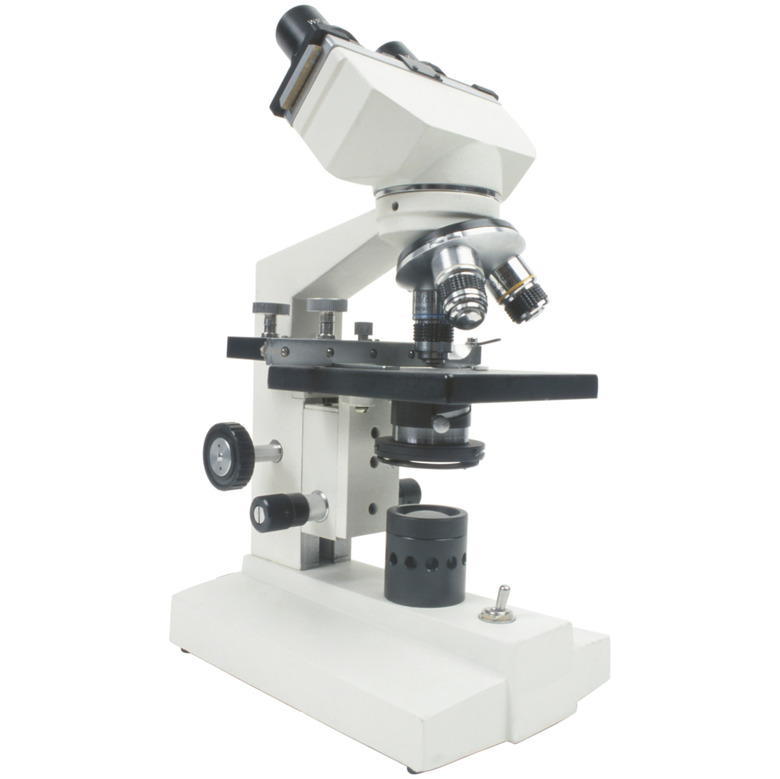
Microscopes are a staple of medical offices, laboratories and science classrooms everywhere. There are several different kinds of microscopes, but the most common type in use is the bright light microscope. It is also known as a bright field microscope. The bright field microscope, despite being one of the simplest and least expensive types of microscope, still has precision components that work together to magnify specimens.
Light Source
Light Source
A light source is necessary to illuminate a specimen. Light may be provided by an external source, although most models have an enclosed incandescent bulb powered either by battery or household current. Some models have an adjustable iris diaphragm that allows the user to adjust the intensity and brightness of the light. The light shines through a condenser, which can be raised and lowered to focus the light beam onto the specimen. Intensity and focus are dependent on the type of specimen and the magnification used.
Stage
Stage
The specimen is placed on the stage for examination. The stage is located above the light source and below the lenses. Specimens are mounted between two small glass plates, called slides. Specimens generally work better if they are thin and transparent or semi-transparent; and sometimes need to be stained to increase contrast. Common specimens include tissue sections, plant sections and various fluids such as blood or pond water.
Lenses
Lenses
The bright light microscope has two sets of lenses, the objective lens and the ocular lens. The objective lens is directly above the stage, and provides the primary magnification. There are often several objective lenses of different powers on a rotating disc. The ocular lens is located at the top of microscope, closest to the user's eyes. It provides the fine tuning necessary to fully focus on the specimen. The light shining up through the specimen and into the lenses creates the image seen by the user.
Focus
Focus
The lenses must be focused in order to obtain a sharp view of the specimen. There are two knobs on the body of the microscope that control focus: the coarse adjustment knob and the fine adjustment knob. Turning the knobs adjusts the distance between the stage and the lens. The coarse adjustment knob is used to bring the specimen into initial focus — visible but not sharp. The fine adjustment knob is then turned to bring the specimen into sharp focus.
Cite This Article
MLA
J.D., Randall Pierce. "How Do Bright Light Microscopes Work?" sciencing.com, https://www.sciencing.com/bright-light-microscopes-work-12122236/. 24 April 2017.
APA
J.D., Randall Pierce. (2017, April 24). How Do Bright Light Microscopes Work?. sciencing.com. Retrieved from https://www.sciencing.com/bright-light-microscopes-work-12122236/
Chicago
J.D., Randall Pierce. How Do Bright Light Microscopes Work? last modified March 24, 2022. https://www.sciencing.com/bright-light-microscopes-work-12122236/
