Cell Structure Definitions
Cells, generally speaking, are similar-to-identical units that make up a whole. Prison blocks and beehives, for example, are made up mostly of cells. As applied to biological systems, the term was likely coined by the 17th-century scientist Robert Hooke, inventor of the compound microscope and pioneer in a remarkable number of scientific endeavors. A cell, as described today, is the smallest unit of a living thing that retains the characteristics of life itself. In other words, individual cells not only contain genetic information, but they also use and transform energy, host chemical reactions, maintain equilibrium and so on. More colloquially, cells are typically and appropriately called "the building blocks of life."
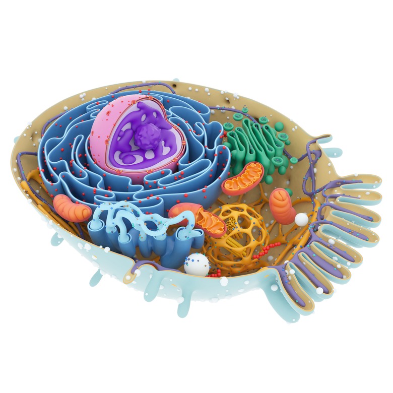
The essential characteristics of a cell include a cell membrane to separate and protect the cell contents from the rest of the world; cytoplasm, or a liquid-like substance in the cell interior in which metabolic processes occur; and genetic material (deoxyribonucleic acid, or DNA). This essentially describes a prokaryotic, or bacterial, cell in its entirety. More complex organisms, however, called eukaryotes – including animals, plants and fungi – feature a variety of other cell structures as well, all of them evolved in accordance with the needs of highly specialized living things. These structures are called organelles. Organelles are to eukaryotic cells what your own organs (stomach, liver, lungs and so on) are to your body as a whole.
Basic Cell Structure
Basic Cell Structure
Cells, structurally, are units of organization. They are formally classified on the basis of where they get their energy. Prokaryotes include two of the six taxonomic kingdoms, Archaebacteria and Monera; all of these species are single-celled and most are bacteria, and they date back an astonishing 3.5 billion years or so (about 80 percent of the estimated age of the Earth itself). Eukaryotes are a "mere" 1.5 billion years old and include Animalia, Plantae, Fungae and Protista. Most eukaryotes are multicellular, although some (e.g., yeast) are not.
Prokaryotic cells, at an absolute minimum, feature an agglomeration of genetic material in the form of DNA inside an enclosure bounded by a cell membrane, also called a plasma membrane. Within this enclosure is, also, is cytoplasm, which in prokaryotes has the consistency of wet asphalt; in eukaryotes, it is much more fluid. In addition, many prokaryotes also have a cell wall outside the cell membrane to serve as a protective layer (as you'll see, the cell membrane serves a variety of functions). Notably, plant cells, which are eukaryotic, also include cell walls. But prokaryotic cells do not include organelles, and this is the primary structural distinction. Even if one chooses to view the distinction as a metabolic one, this is still linked to the respective structural properties.
Some prokaryotes have flagella, which are whip-like polypeptides used for propulsion. Some also have pili, which are hair-like projections used for adhesive purposes. Bacteria also come in multiple shapes: Cocci are round (like the meningococci, which can cause meningitis in humans), baccilli (rods, like the species that cause anthrax), and spirilla or spirochetes (helical bacteria, like those responsible for causing syphilis).
What about viruses? These are merely tiny bits of genetic material, which can be DNA or RNA (ribonucleic acid), surrounded by a protein coat. Viruses are unable to reproduce on their own, and must therefore must infect cells and "hijack" their reproductive apparatus in order to propagate copies of themselves. Antbiotics, as a result, target all manner of bacteria but are ineffective against viruses. Antiviral drugs exist, with newer and more effective ones being introduced all the time, but their mechanisms of action are completely different from those of antibiotics, which usually target either cell walls or metabolic enzymes particular to prokaryotic cells.
The Cell Membrane
The Cell Membrane
The cell membrane is a multifaceted wonder of biology. Its most obvious job is to serve as a container for the contents of the cell and provide a barrier to the insults of the extra-cellular environment. This, however, only describes a small part of its function. The cell membrane is not a passive partition but a highly dynamic assembly of gates and channels that help ensure the maintenance of a cell's internal environment (that is, its equilibrium or homeostasis) by selectively allowing molecules into and out of the cell as required.
The membrane is actually a double membrane, with two layers facing each other in a mirror-image fashion. This is called the phospholipid bilayer, and each layer consists of a "sheet" of phospholipid molecules, or more properly, glycerophospholipid molecules. These are elongated molecules consisting of polar phosphate "heads" that face away from the center of the bilayer (that is, toward the cytoplasm and the cell exterior) and nonpolar "tails" consisting of a pair of fatty acids; these two acids and the phosphate are attached to opposite sides of a three-carbon glycerol molecule. Because of the asymmetrical charge distribution on phosphate groups and the lack of charge asymmetry of fatty acids, phospholipids placed in solution actually assemble themselves spontaneously into this sort of bilayer, so it is energetically efficient.
Substances can traverse the membrane in a variety of ways. One is simple diffusion, which sees small molecules such as oxygen and carbon dioxide move through the membrane from regions of higher concentration to areas of lower concentration. Facilitated diffusion, osmosis and active transport also help maintain a steady supply of nutrients coming into the cell and metabolic waste products exiting.
The Nucleus
The Nucleus
The nucleus is the site of DNA storage in eukaryotic cells. (Recall that prokaryotes lack nuclei because they lack membrane-bound organelles of any sort.) Like the plasma membrane, the nuclear membrane, also called a nuclear envelope, is a double-layered phospholipid barrier.
Within the nucleus, the genetic material of a cell is arranged into distinct bodies called chromosomes. The number of chromosomes an organism has varies from species to species; humans have 23 pairs, including 22 pairs of "normal" chromosomes, called autosomes, and one pair of sex chromosomes. The DNA of individual chromosomes is arranged in sequences called genes; each gene carries the genetic code for a particular protein product, be it an enzyme, a contributor to eye color or a component of skeletal muscle.
When a cell undergoes division, its nucleus divides in a distinct way, owing to the replication of the chromosomes within it. This reproductive process is called mitosis, and the cleavage of the nucleus is known as cytokinesis.
Ribosomes
Ribosomes
Ribosomes are the site of protein synthesis in cells. These organelles are made almost entirely from a type of RNA fittingly called ribosomal RNA, or rRNA. These ribosomes, which are found throughout the cell cytoplasm, include one large subunit and one small subunit.
Perhaps the easiest way to envision ribosomes is as tiny assembly lines. When it is time to manufacture a given protein product, messenger RNA (mRNA) transcribed in the nucleus from DNA makes its way to the portion of ribosomes where the mRNA code is translated into amino acids, the building blocks of all proteins. Specifically, the four different nitrogenous bases of mRNA can be arranged in 64 different ways into groups of three (4 raised to the third power is 64), and each of these "triplets" codes for an amino acid. Because there are only 20 amino acids in the human body, some amino acids are derived from more than one triplet code.
When the mRNA is being translated, yet another type of RNA, transfer RNA (tRNA) carries whatever amino acid has been summoned by the code to the ribosomal site of synthesis, where the amino acid is attached to the end of the protein-in-progress. Once the protein, which can be anywhere from dozens to many hundreds of amino acids long, is complete, it is released from the ribosome and transported to wherever it is needed.
Mitochondria and Chloroplasts
Mitochondria and Chloroplasts
Mitochondria are the "power plants" of animal cells, and chloroplasts are their analogs in plant cells. Mitochondria, believed to have originated as free-standing bacteria before becoming incorporated into the structures that became eukaryotic cells, are the site of aerobic metabolism, which requires oxygen to extract energy in the form of adenosine triphosphate (ATP) from glucose. The mitochondria receives pyruvate molecules derived from oxygen-independent glucose breakdown in the cytoplasm; in the matrix (interior) of the mitochondria, the pyruvate is subjected to the Krebs cycle, also called the citric-acid cycle or the tricarboxylic acid (TCA) cycle. The Krebs cycle generates a build-up of high-energy proton carriers and serves as set-up for the aerobic reactions called the electron transport chain, which occurs nearby on the mitochondrial membrane, which is yet another lipid bilayer. These reactions generate far more energy in the form of ATP than glycolysis can; without mitochondria, animal life could not have evolved on Earth owing to the prodigious energy requirements of "higher" organisms.
Chloroplasts are what give plants their green color, as they contain a pigment called chlorophyll. Whereas mitochondria breaks down glucose products, chloroplasts actually use the energy from sunlight to build glucose from carbon dioxide and water. The plant then uses some of this fuel for its own needs, but most of it, along with the oxygen liberated in glucose synthesis, reaches the ecosystem and is used by animals, which cannot make their own food. Without abundant plant life on Earth, animals could not survive; the converse is true, as animal metabolism generates sufficient carbon dioxide for plants to use.
The Cytoskeleton
The Cytoskeleton
The cytoskeleton, as its name suggests, provides structural support to a cell in the same way your own bony skeleton provides a stable scaffolding for your organs and tissues. The cytoskeleton consists of three components: microfilaments, intermediate fibers and microtubules, in order from smallest to largest. Microfilaments and microtubules can be assembled and disassembled according to the needs of cell at a given time, whereas intermediate filaments tend to be more permanent.
In addition to fixing organelles in place much like the guide wires attached to tall communication towers keep these fixed to the ground, the cytoskeleton assists in moving things within a cell. This can be in the form of serving as anchor points for flagella, as some microtubules do; alternatively, some microtubules provide the actual conduit (pathway) for things to move along. Thus the cytoskeleton can be both motor and highway, depending on the specific type.
Other Organelles
Other Organelles
Other important organelles include Golgi bodies, which look like stacks of pancakes on microscopic examination and serve as sites of protein storage and secretion, and the endoplasmic reticulum, which moves protein products along from one portion of the cell to another. Endoplasmic reticulum comes in smooth and rough forms; the latter are so named because they are studded with ribosomes. Golgi bodies give rise to vesicles that break off the edges of the "pancakes" and contain proteins; if these can be regarded as shipping containers, then the endoplasmic reticulum that receives these bodies is like a highway or railroad system.
Lysosomes are also important in the upkeep of cells. These are also vesicles, but they contain specific digestive enzymes that can lyse (dissolve) either the metabolic waste products of cells or chemicals that are not supposed to be there at all but have somehow breached the cell membrane.
Cite This Article
MLA
Beck, Kevin. "Cell Structure Definitions" sciencing.com, https://www.sciencing.com/cell-structure-definitions-5043056/. 24 August 2018.
APA
Beck, Kevin. (2018, August 24). Cell Structure Definitions. sciencing.com. Retrieved from https://www.sciencing.com/cell-structure-definitions-5043056/
Chicago
Beck, Kevin. Cell Structure Definitions last modified August 30, 2022. https://www.sciencing.com/cell-structure-definitions-5043056/
