Cell Wall: Definition, Structure & Function (With Diagram)

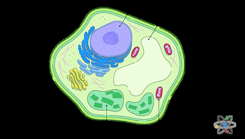
The cell wall is an additional layer of protection on top of the cell membrane. You can find cell walls in both prokaryotes and eukaryotes, and they are most common in plants, algae, fungi and bacteria.
However, animals and protozoans do not have this type of structure. Cell walls tend to be rigid structures that help maintain the shape of the cell.
What Is the Function of a Cell Wall?
What Is the Function of a Cell Wall?
The cell wall has several functions, including the maintenance of the cell structure and shape. The wall is rigid, so it protects the cell and its contents.
For example, the cell wall can keep pathogens like plant viruses from entering. In addition to the mechanical support, the wall acts as a framework that can prevent the cell from expanding or growing too quickly. Proteins, cellulose fibers, polysaccharides and other structural components help the wall maintain the shape of the cell.
The cell wall also plays an important role in transport. Since the wall is a semi-permeable membrane, it allows certain substances to pass through, such as proteins. This allows the wall to regulate diffusion in the cell and control what enters or leaves.
Additionally, the semi-permeable membrane helps communication among cells by allowing signaling molecules to pass through the pores.
What Makes Up the Plant Cell Wall?
What Makes Up the Plant Cell Wall?
A plant cell wall consists primarily of carbohydrates, like pectins, cellulose and hemicellulose. It also has structural proteins in smaller amounts and some minerals such as silicon. All of these components are vital parts of the cell wall.
Cellulose is a complex carbohydrate and consists of thousands of glucose monomers that form long chains. These chains come together and form cellulose microfibrils, which are several nanometers in diameter. The microfibrils help control the growth of the cell by limiting or allowing its expansion.
Turgor Pressure
Turgor Pressure
One of the main reasons for having a wall in a plant cell is that it can withstand turgor pressure, and this is where cellulose plays a crucial role. Turgor pressure is a force created by the inside of the cell pushing out. Cellulose microfibrils form a matrix with the proteins, hemicelluloses and pectins to provide the strong framework that can resist turgor pressure.
Both hemicelluloses and pectins are branched polysaccharides. Hemicelluloses have hydrogen bonds connecting them to the cellulose microfibrils, while pectins trap water molecules to create a gel. Hemicelluloses increase the strength of the matrix, and pectins help prevent compression.
Proteins in the Cell Wall
Proteins in the Cell Wall
The proteins in the cell wall serve different functions. Some of them provide structural support. Others are enzymes, which are a type of protein that can speed up chemical reactions.
The enzymes help the formation of and normal modifications that occur to maintain the plant's cell wall. They also play a part in fruit ripening and leaf color changes.
If you have ever made your own jam or jelly, then you have seen the same types of pectins found in cell walls in action. Pectin is the ingredient that cooks add to thicken fruit juices. They often use the pectins naturally found in apples or berries to make their jams or jellies.
 Sciencing
Structure of the Plant Cell Wall
Structure of the Plant Cell Wall
Plant cell walls are three-layered structures with a middle lamella, primary cell wall and secondary cell wall. The middle lamella is the outermost layer and helps with cell-to-cell junctions while holding adjacent cells together (in other words, it sits between and holds together the cell walls of two cells; this is why it's called the middle lamella, even though it is the outermost layer).
The middle lamella acts like glue or cement for plant cells because it contains pectins. During cell division, the middle lamella is the first to form.
Primary Cell Wall
Primary Cell Wall
The primary cell wall develops when the cell grows, so it tends to be thin and flexible. It forms between the middle lamella and the _plasma membrane_.
It consists of cellulose microfibrils with hemicelluloses and pectins. This layer allows the cell to grow over time but does not overly restrict the cell's growth.
Secondary Cell Wall
Secondary Cell Wall
The secondary cell wall is thicker and more rigid, so it provides more protection for the plant. It exists between the primary cell wall and the plasma membrane. Often, the primary cell wall actually helps create this secondary wall after the cell finishes growing.
Secondary cell walls consist of cellulose, hemicelluloses and lignin. Lignin is a polymer of aromatic alcohol that provides additional support for the plant. It helps protect the plant from attacks by insects or pathogens. Lignin also helps with water transport in the cells.
Difference Between Primary and Secondary Cell Walls in Plants
Difference Between Primary and Secondary Cell Walls in Plants
When you compare the composition and thickness of primary and secondary cell walls in plants, it is easy to see the differences.
First, primary walls have equal amounts of cellulose, pectins and hemicelluloses. However, secondary cell walls do not have any pectin and have more cellulose. Second, the cellulose microfibrils in primary cells walls look random, but they are organized in secondary walls.
Although scientists have discovered many aspects of how cell walls function in plants, some areas still need more research.
For example, they are still learning more about the actual genes involved in the biosynthesis of the cell wall. Researchers estimate that about 2,000 genes take part in the process. Another important area of study is how gene regulation works in the plant cells and how it affects the wall.
The Structure of Fungal and Algal Cell Walls
The Structure of Fungal and Algal Cell Walls
Similar to plants, the cell walls of fungi consist of carbohydrates. However, while fungi have cells with chitin and other carbohydrates, they do not have cellulose like plants do.
Their cell walls also have:
- Enzymes
- Glucans
- Pigments
- Waxes
- Other substances
It is important to note that not all fungi have cell walls, but many of them do. In fungi, the cell wall sits outside the plasma membrane. Chitin makes up most of the cell wall, and it is the same material that gives insects their strong exoskeletons.
Fungal Cell Walls
Fungal Cell Walls
In general, fungi with cell walls have three layers: chitin, glucans and proteins.
As the innermost layer, chitin is fibrous and made up of polysaccharides. It helps make the fungi cell walls rigid and strong. Next, there is a layer of glucans, which are glucose polymers, crosslinking with chitin. The glucans also help the fungi maintain their cell wall rigidity.
Finally, there is a layer of proteins called the mannoproteins or mannans, which have a high level of mannose sugar. The cell wall also has enzymes and structural proteins.
Different components of the fungal cell wall can serve different purposes. For instance, enzymes can help with digestion of organic materials, while other proteins may help with adhesion in the environment.
Cell Walls in Algae
Cell Walls in Algae
The cell walls in algae consist of polysaccharides, like cellulose, or glycoproteins. Some algae have both polysaccharides and glycoproteins in their cell walls. In addition, algal cell walls have mannans, xylans, alginic acid and sulfonated polysaccharides. The cell walls among different types of algae can vary greatly.
Mannans are proteins that make microfibrils in some green and red algae. Xylans are complex polysaccharides and sometimes replace cellulose in algae. Alginic acid is another type of polysaccharide often found in brown algae. However, most algae have sulfonated polysaccharides.
Diatoms are a type of algae that live in water and soil. They are unique because their cell walls are made of silica. Researchers are still investigating how **diatoms** form their cell walls and which proteins make up the process.
Nevertheless, they have determined that diatoms form their mineral-rich walls internally and move them outside the cell. This process, called exocytosis, is complex and involves multiple proteins.
Bacterial Cell Walls
Bacterial Cell Walls
A bacterial cell wall has peptidoglycans. Peptidoglycan or murein is a unique molecule that consists of sugars and amino acids in a mesh layer, and it helps the cell maintain its shape and structure.
The cell wall in bacteria exists outside of the plasma membrane. Not only does the wall help configure the shape of the cell, but it also helps prevent the cell from bursting and spilling all of its content.
Gram-Positive and Gram-Negative Bacteria
Gram-Positive and Gram-Negative Bacteria
In general, you can divide bacteria into gram-positive or gram-negative categories, and each type has a slightly different cell wall. Gram-positive bacteria can stain blue or violet during a Gram staining test, which uses dyes to react with the peptidoglycans in the cell wall.
On the other hand, gram-negative bacteria cannot be stained blue or violet with this type of test. Today, microbiologists still use Gram staining to identify the type of bacteria. It is important to note that both gram-positive and gram-negative bacteria have peptidoglycans, but an extra outer membrane prevents the staining of gram-negative bacteria.
Gram-positive bacteria have thick cell walls made from layers of peptidoglycans. Gram-positive bacteria have one plasma membrane surrounded by this cell wall. However, gram-negative bacteria have thin cell walls of peptidoglycans that are not enough to protect them.
This is why gram-negative bacteria have an additional layer of lipopolysaccharides (LPS) that serve as an endotoxin. Gram-negative bacteria have an inner and outer plasma membrane, and the thin cell walls are in between the membranes.
Antibiotics and Bacteria
Antibiotics and Bacteria
The differences between human and bacterial cells make it possible to use antibiotics in your body without killing all of your cells. Since people do not have cell walls, medications like antibiotics can target cell walls in bacteria. The composition of the cell wall plays a role in how some antibiotics work.
For example, penicillin, a common beta-lactam antibiotic, can affect the enzyme that forms the links between peptidoglycan strands in bacteria. This helps to destroy the protective cell wall and stops the bacteria from growing. Unfortunately, antibiotics can kill both helpful and harmful bacteria in the body.
Another group of antibiotics called glycopeptides targets the synthesis of cell walls by stopping peptidoglycans from forming. Examples of glycopeptide antibiotics include vancomycin and teicoplanin.
Antibiotic Resistance
Antibiotic Resistance
Antibiotic resistance happens when bacteria change, which makes the drugs less effective. Since the resistant bacteria survive, they can reproduce and multiply. Bacteria become resistant to antibiotics in different ways.
For instance, they can change their cell walls. They can move the antibiotic out of their cells, or they can share genetic information that includes resistance to the drugs.
One way some bacteria resist beta-lactam antibiotics like penicillin is to make an enzyme called beta-lactamase. The enzyme attacks the beta-lactam ring, which is a core component of the drug, and consists of carbon, hydrogen, nitrogen and oxygen. However, drug manufacturers try to prevent this resistance by adding beta-lactamase inhibitors.
Cell Walls Matter
Cell Walls Matter
Cell walls offer protection, support and structural help for plants, algae, fungi and bacteria. Although there are major differences among the cell walls of prokaryotes and eukaryotes, most organisms have their cell walls outside of the plasma membranes.
Another similarity is that most cell walls provide rigidity and strength that help the cells maintain their shape. Protection from pathogens or predators is also something that many cell walls among different organisms have in common. Many organisms have cell walls made up of proteins and sugars.
Understanding the cell walls of prokaryotes and eukaryotes can help people in a variety of ways. From better medications to stronger crops, learning more about the cell wall offers a lot of potential benefits.
References
- Florida State University: Molecular Expressions: Plant Cell Wall
- National Institute of General Medical Sciences: Studying Cells
- BBC: GCSE Edexcel: Cell Structure
- BMC Biology: An Inside-out Origin for the Eukaryotic Cell
- New World Encyclopedia: Cell Wall
- Frontiers in Marine Science: Understanding Diatom Cell Wall Silicification — Moving Forward
- CDC: Antibiotic Resistance Questions and Answers
Cite This Article
MLA
Bandoim, Lana. "Cell Wall: Definition, Structure & Function (With Diagram)" sciencing.com, https://www.sciencing.com/cell-wall-definition-structure-function-with-diagram-13717284/. 15 March 2019.
APA
Bandoim, Lana. (2019, March 15). Cell Wall: Definition, Structure & Function (With Diagram). sciencing.com. Retrieved from https://www.sciencing.com/cell-wall-definition-structure-function-with-diagram-13717284/
Chicago
Bandoim, Lana. Cell Wall: Definition, Structure & Function (With Diagram) last modified March 24, 2022. https://www.sciencing.com/cell-wall-definition-structure-function-with-diagram-13717284/