Centrosome: Definition, Structure & Function (With Diagram)

The centrosome ("middle body") is a structure found in the cells of most plants and animals. It is from this organelle that the protein structures known as microtubules form and extend.
These microtubules emerge from the microtubule organizing center (MTOC), and are integral to a number of eukaryotic cell functions and processes throughout a cell's lifetime. They are perhaps best known for their important role in the process of cell division, which includes mitosis (the division of a cell's nuclear material into daughter nuclei) followed in short order by cytokinesis (the division of an entire cell into daughter cells).
This division process is mediated by the centrioles of centrosomes.
The Structure of the Centriole
The Structure of the Centriole
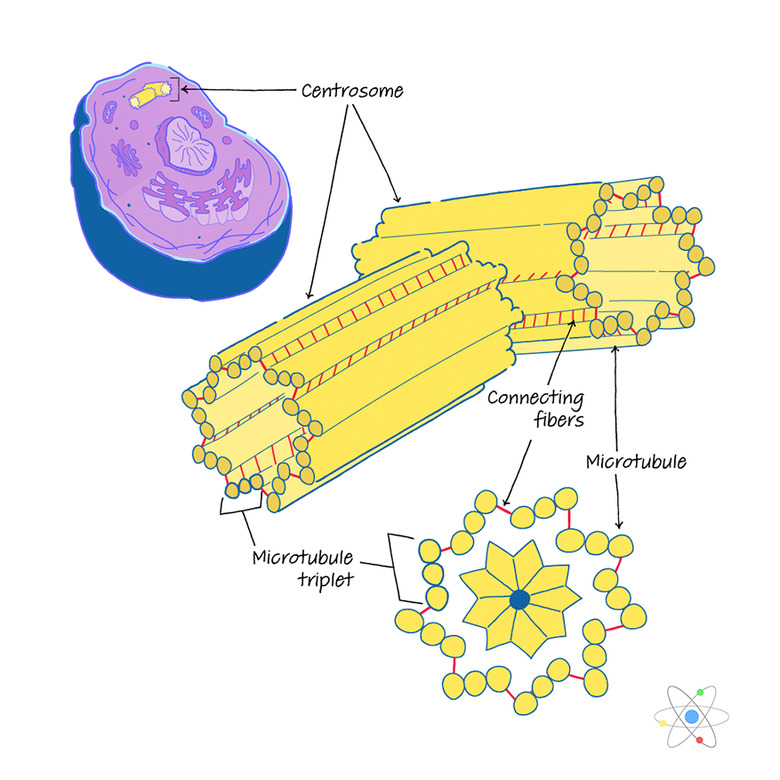
Centrosomes are structures that contain the centrioles, which give rise to the microtubules that function as the mitotic spindle. That's a lot to envision, so a look at each of these in terms gives a clearer idea of the physical set-up of centrosomes.
During interphase, which is the period during which a cell is not actively dividing, each cell contains one centrosome that includes a pair of centrioles. Each of these centrioles consists of nine microtubule triplets in a cylindrical arrangement; in other words, a single centriole includes a total of 27 microtubules running from end to end. The two centrioles are oriented at right angles to one another. The triplets themselves resemble tiny parallel pipes that are in a line.
Read more about what happens in interphase.
- If you were to look at a cross-section of a centriole, you'd see a circular formation consisting of **nine groups..**.
- ...and each of these groups has a line of three smaller circles, with these lines of smaller circles angled toward the middle of the circular formation.
Also during interphase, all of a cell's basic components are replicated, including the centrosome and its pair of centrioles. Initially, the two centrosomes, or pairs of centrioles, remain in close physical proximity. Once mitosis is fully underway, the two centrioles migrate toward opposite ends of the cell that is preparing to split into two daughter cells.
- Between the centrioles and the cellular matrix in which they are created and reside, over 100 distinct proteins have a function in the structure of the centrosome. This matrix is known as the pericentriolar material, or PCM.
**Centrosome vs. Centromere:** Neither "centrosome" or "centriole" should be confused with the centromere, which is the physical junction between the sister chromatids of a chromosome that is preparing to divide as part of mitosis.)
Microtubules, as noted, have a number of different functions in cells, but their main purpose in cell division is to serve as the spindle fibers that help control and carry out the separation of cellular components during the division process.
The Centrosome as Part of the Cytoskeleton
The Centrosome as Part of the Cytoskeleton
In addition to taking part in mitosis, the centrosome plays a vital structural role in the cell by generating the microtubules that form the cytoskeleton, which is what gives cells their shape and integrity.
Although it is perhaps tempting to imagine cells as fragile, gelatinous globs that are little more than roundish containers, every cell is extremely dynamic, including its membrane, which carefully controls what substances may or may not pass into and outside of the cell.
- If the microtubules that participate in cell division by forming the spindle are like levers that control where parts of the cell go, then the ones that make up the static cytoskeleton are like scaffolding.
Read more about the main function of microtubules cells.
Their purpose is similar to that of your own body's skeleton, which gives the rest of you your general physical shape and functions as a rack of sorts that holds your other important physical components – your organs, muscles and tissues.
**Cytoskeleton Arrangement and Composition:** The microtubules forming the cytoskeleton are threaded across the cytoplasm of the cell's interior, forming a series of braces between the cell's boundary and its nucleus close to the center. These tubules in turn consist of monomeric units made of a protein called tubulin.
This tubulin, like many proteins in nature, comes in a variety of subtypes; the most common found in microtubules are:
- alpha-tubulin
- beta-tubulin
Only in the presence of a centrosome do these monomers spontaneously form themselves into microtubules, in much the same way, perhaps, as eggs, sugar and chocolate only form themselves into cookies in the presence of a human-staffed kitchen.
In addition, proteins called dyneins and kinesins take part in mitosis; these help orient the ends of the microtubules to their correct locations along or near the soon-to-divide chromosomes, which line up along the metaphase plate.
**Importance of Centrosomes:** It is not yet known how exactly the duplication of centrosomes during interphase occurs. Also, it is notable that while centrosomes and centrioles do appear in most plant cells, mitosis can occur in plants in the absence of these structures. In fact, in some animal cells, mitosis can function even when the centrioles have been purposefully destroyed, but this generally results in an unusually high number of replication errors.
It is therefore believed that centrosomes help impart a degree of control over the entire process, and biochemists are striving to elucidate the mechanisms of this because those are likely important in the genesis and progression of cancers and other disorders that are contingent on cell replication and division.
 Dana Chen | Sciencing
Role of the Centrosome in Cell Division
Role of the Centrosome in Cell Division
Cell division is a crucial component of cell biology. Centrosomes play a major role this process.
Remember that the two centrioles of a single centrosome are oriented at right angles to each other, meaning that the microtubules in these centrioles will be arrayed in one of two mutually perpendicular directions. Also recall that the two centrosomes in an as-yet-not-quite-dividing cell lie on opposite ends of the interphase cell.
An implication of this geometry is that when the spindle fibers of mitosis begin to form, they extend from each side (or "pole") of the cell toward its center, where cell division is ultimately most evident, and they also extend or "fan" outward in a range of directions from each centrosome itself.
Try holding your closed fists held slightly apart, and then slowly open them while extending your newly visible fingers toward each other; this offers a general picture of what unfolds at the centrosomes as mitosis proceeds.
Mitosis itself includes four phases (sometimes listed as five). In order, these are:
1. Prophase 2. Metaphase 3. Anaphase 4. Telophase
Some sources also include prometaphase between prophase and metaphase. As mitosis gets underway, the microtubules growing from the nascent mitotic spindle at each pole move toward the center of the cell, where the replicated chromosomes arranged in pairs are lined up along the so-called metaphase plate (an invisible line along which the cleavage of the nucleus occurs).
These ranging ends of the spindle fibers wind up in one of three places: on the kinetochore of each chromosome pair, which is the structure at which chromosomes actually separate; on the arms of the chromosomes; and in the cytoplasm itself well on the other side of the cell, closer to the opposing centrosome than to these fibers' point of origin.
**Spindle Fibers in Operation:** The range of anchor points of the ends of the spindle fibers attests to the elegance and complexity of the mitotic process. It is a "tug-of-war" of sorts, but one that must be extremely well coordinated, so that division "runs through" the exact middle of every chromosome pair to ensure that each daughter cell receives exactly one chromosome from each pair.
The spindle fibers therefore do some "pushing" as well as a great deal of "pulling" to make sure cell division is not only forceful but accurate. The microtubules take part in the division of the nucleus alone, but also participate in the division of the entire cell (i.e., cytokinesis) and the re-enclosure of each new daughter cell in its own cell membrane.
One way to perhaps imagine all of this: Cells do not have muscles, but the microtubules are about as close as cell components get.
Centriole Replication
Centriole Replication
As stated, the centrosomes of cells replicate during interphase, the comparatively long part of the cell cycle between mitotic divisions. The replication of centrioles in centrosomes is not fully conservative, meaning that the two daughter centrioles formed are not entirely identical, as would occur in a conservative process. Instead, centriole replication is semiconservative.
While the exact mechanism of centrosome replication during the **S phase (synthesis phase)** of cell interphase remains to be fully understood, scientists have realized that when a centriole divides, one of the resultant centrioles retains the characteristics of the "mother" and can generate operational microtubules.
This centriole has "stem-cell-like" properties, whereas the other, the "daughter," becomes fully differentiated. Each dividing cell has one mother-daughter centriole pair at each pole, so **each new daughter cell, as you might expect, contains one mother centriole and one daughter centriole in each pair.** During the interphase that soon follows, this centriole will divide to again create two mother centriole-daughter centriole pairs.
**Centrioles in Differentiated Structures:** The subtle differences in function between the right-angled centrioles in each pair become evident when, for example, the mother centriole becomes attached to the interior of the cell's plasma membrane to form a structure called a basal body. This body is usually part of a cilium, or hair-like multi-microtubule extension, that is not motile; that is, it doesn't move.
Some cilia (the plural of "cilium") form flagella (singular "flagellum") that do move, often propelling entire cells along while in other cases serving as miniature brooms of a sort that clear debris from the region of the flagellum.
While biologists have a lot to learn about the precise dynamics of centrosomes, cancer provides a window into what goes wrong with centrosomes in instances of abnormal cell division. Researchers have observed, for example, that cancer cells often contain unusual numbers of centrosomes instead of the expected one or two, and certain anti-cancer drugs (for example, Taxol and vincristine) exert their effects by interfering with microtubule assembly.
Role in Cilia Formation
Role in Cilia Formation
A flagellum is an assortment of microtubules that allows for locomotion, as in the case of a sperm cell. A flagellum originates from a single basal body on the inner surface of the plasma membrane. Thus, a sperm cell contains a single centriole pair.
Because a sperm cell's ultimate fate is to fuse with an egg cell, and an egg cell lacks a basal body, it is the sperm that ensures that a newly formed zygote (the product of egg-sperm joining and the first step in the generation of a new organism in reproduction) will be able to divide, since **the centriole includes instructions and components needed for the division process.**
Some organisms have cilia on certain cells. This includes some of the cells of your own respiratory tract. The epithelium (surface cells; your skin is a sort of epithelium) that lines your lungs forms a number of connected basal bodies, which is what a cilium actually is. The tubular extensions of these ciliated cells function to move along mucus and particulate matter and therefore protect the interior of the lungs.
Cite This Article
MLA
Beck, Kevin. "Centrosome: Definition, Structure & Function (With Diagram)" sciencing.com, https://www.sciencing.com/centrosome-definition-structure-function-with-diagram-13717286/. 30 July 2019.
APA
Beck, Kevin. (2019, July 30). Centrosome: Definition, Structure & Function (With Diagram). sciencing.com. Retrieved from https://www.sciencing.com/centrosome-definition-structure-function-with-diagram-13717286/
Chicago
Beck, Kevin. Centrosome: Definition, Structure & Function (With Diagram) last modified March 24, 2022. https://www.sciencing.com/centrosome-definition-structure-function-with-diagram-13717286/