What Is The Chromatin's Function?
Most everyday people are familiar with deoxyribonucleic acid, or DNA, be it from following crime shows or exposure to basic genetics. Similarly, the typical high-school student has most likely heard the term "chromosome" in some context, and recognize that these, too, relate to how living things reproduce and dispatch heritable traits to their offspring. But the entity that functionally joins DNA and chromosomes, called chromatin, is far more obscure.
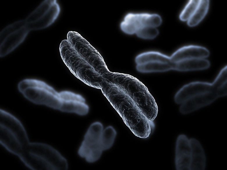
DNA is a molecule possessed by all living things that is divided into genes, the biochemical codes for making specific protein products. Chromosomes are very long strips of DNA bonded to proteins that ensure that the chromosomes can change their shape dramatically depending the life stage of the cell in which they sit. Chromatin is simply the material from which chromosomes are made; put differently, chromosomes are nothing more than discrete pieces of chromatin.
An Overview of DNA
An Overview of DNA
DNA is one of two nucleic acids found in nature, the other being ribonucleic acid (RNA). In prokaryotes, almost all of which are bacteria, the DNA sits in a loose cluster in the bacterial cell's cytoplasm (the semi-liquid, protein-rich interior of cells) rather than in a nucleus, as these simple organisms lack internal membrane-bound structures. In eukaryotes (i.e., animals, plants and fungi), the DNA lies inside the cell nucleus.
DNA is a polymer assembled from subunits (monomers) called nucleotides. Each nucleotide in turn includes a pentose, or five-carbon, sugar (deoxyribose in DNA, ribose in RNA), a phosphate group and a nitrogenous base. Each nucleotide boasts one of four different nitrogenous bases available to it; in DNA, these are adenine (A), cytosine (C), guanine (G) and thymine (T). In RNA, the first three are present, but uracil, or U, is substituted for thymine. Because it is the bases that distinguish the four DNA nucleotides from each other, it is their sequence that determines the uniqueness of every individual organism's DNA as a whole; every one of the billions of people on Earth has a genetic code, determined by DNA, that no one else has, unless he or she has an identical twin.
DNA is in a double-stranded, helical formation in its most chemically stable form. The two strands are bound at each nucleotide by their respective bases. A pairs with and only with T, while C pairs with and only with G. Because of this specific base pairing, each DNA strand in a whole molecule is complementary to the other. DNA therefore resembles a ladder that has been twisted many times at each of its ends in opposite directions.
When DNA makes copies of itself, this process is called replication. DNA's main job is to send the code for making proteins to the parts of the cell it needs to go. It does this by relying on a biochemical courier that, as luck would have it, DNA also happens to create. Single strands of DNA are used to build a molecule called messenger RNA, or mRNA. This process is called transcription, and the result is a mRNA molecule with a nucleotide base sequence that is complementary to the DNA strand from which is was constructed (called the template) except that U appears in the mRNA strand in place of T. This mRNA then migrates toward structures in the cytoplasm called ribosomes, which use the mRNA instructions to build specific proteins.
Chromatin: Structure
Chromatin: Structure
In life, almost all DNA exists in the form of chromatin. Chromatin is DNA bound to proteins called histones. These histone proteins, which make up about two-thirds of the mass of chromatin, include four subunits arranged in pairs, with the pairs all grouped together to form an eight-subunit structure called an octamer. These histone octamers bind to DNA, with DNA winding around the octamers like thread around a spool. Each octamer has a little less than two full turns of DNA wrapped around it – about 146 base pairs in total. The entire result of these interactions – the histone octamer and the DNA surrounding it – is called a nucleosome.
The reason for what at a glance seems to be excess protein baggage is straightforward: Histones, or more specifically nucleosomes, are what permit DNA to be compacted and folded to the extreme degree required for all of a single complete copy of DNA to fit into a cell about one-millionth of a meter wide. Human DNA, of straightened out completely, would measure an astonishing 6 feet – and remember, that is in each one of the trillions of cells in your body.
The histones latched on to DNA to create nucleosomes make chromatin look like a series of beads strung on string under a microscope. This is deceiving, however, because as chromatin exists in living cells, it is far more tightly wound than the coiling around the histone octamers can account for. As it happens, nucleosomes are stacked in layers atop each other, and these stacks in turn are coiled and doubled back in many levels of structural organization. Roughly speaking, the nucleosomes alone increase DNA packing by a factor of about six. The subsequent stacking and coiling of these into a fiber about 30 nanometers wide (30 one-billionths of a meter) increases DNA packing further by a factor of 40. This fiber is wound around a matrix, or core, that affords another 1,000 times the packing efficiency. And fully condensed chromosomes increase this by another 10,000-fold.
The histones, though described above as coming in four different types, are actually very dynamic structures. While the DNA in chromatin retains the same exact structure throughout its lifetime, the histones can have various chemical groups attached to them, including acetyl groups, methyl groups, phosphate groups and more. Because of this, it is likely that histones play a significant role in how the DNA in their midst is ultimately expressed – that is, how many copies of given types of mRNA are transcribed by different genes along the DNA molecule.
The chemical groups attached to histones change which free proteins in the area can bind to given nucleosomes. These bound proteins can change the organization of the nucleosomes in different ways along the length of the chromatin. Also, unmodified histones are highly positively charged, which has them bind tightly with the negatively charged DNA molecule by default; proteins bound to nucleosomes via the histones can lessen histones' positive charge and weaken the histone-DNA bond, allowing the chromatin to "relax" under these conditions. This loosening of chromatin makes the DNA strands more accessible and free to operate, making processes such as replication and transcription easier when the time comes for these to occur.
Chromosomes
Chromosomes
Chromosomes are segments of chromatin in its most active, functional form. Humans have 23 distinct chromosomes, including twenty-two numbered chromosomes and one sex chromosome, either X or Y. Every cell in your body, however, with the exception of gametes, has two copies of every chromosome – 23 from your mother and 23 from your father. The numbered chromosomes vary considerably in size, but chromosomes with the same number, whether they come from your mother or father, all look like same under a microscope. Your two sex chromosomes, however, look different if you are male, because you have one X and one Y. If you are female, you have two X chromosomes. From this, you can see that the male's genetic contribution determines the sex of the offspring; if a male donates an X to the mix, the offspring will have two X chromosomes and be female, whereas if the male donates his Y, the offspring will have one of each sex chromosome and be male.
When chromosomes replicate – that is, when complete copies of all of the DNA and proteins within the chromosomes are made – they then consist of two identical chromatids, called sister chromatids. This happens separately in each copy of your chromosomes, so after replication, you would have a total of four chromatids pertaining to each numbered chromosome: two identical chromatids in your father's copy of, say, chromosome 17 and two identical chromatids in your mother's copy of chromosome 17.
Within each chromosome, the chromatids are joined at a structure of condensed chromatin called the centromere. Despite its somewhat misleading name, the centromere does not necessarily appear halfway along the length of each chromatid; in fact, in most cases it does not. This asymmetry leads to the appearance of shorter "arms" extending in one direction from the centromere, and longer "arms" extending in the other direction. Each short arm is called the p-arm, and the longer arm is known as the q-arm. With the completion of the Human Genome Project in the early part of the 21st century, every specific gene in the body has been localized and mapped onto every arm of every human chromosome.
Cell Division: Meiosis and Mitosis
Cell Division: Meiosis and Mitosis
When everyday cells of the body divide (gametes, discussed shortly, are the exception), they undergo a simple reproduction of the entire cell's DNA, called mitosis. This is what allows cells in the various tissues of the body to grow, be repaired and be replaced when they wear out.
Most of the time, the chromosomes in your body are relaxed and diffuse within the nucleus, and are often referred to simply as chromatin in this state because of the lack of appearance of organization to the extent they can be visualized. When it comes time for the cell to divide, at the end of the G1 phase of the cell cycle, the individual chromosomes replicate (S phase) and begin to condense inside the nucleus.
Thus starts prophase of mitosis (the M phase of the overall cell cycle), the first of four delineated phases of mitosis. Two structures called centrosomes migrate to opposite sides of the cell. These centrosomes form microtubules that extend toward the chromosomes and the middle of the cell, in an outward-radiating way, generating what is called the mitotic spindle. In metaphase, all 46 chromosomes move to the middle of the cell in a line, with this line perpendicular to the line between the chromosomes. Importantly, the chromosomes are aligned so that one sister chromatid is on one side of the soon-to-be-dividing line and one sister chromatid is fully on the other. This assures that when the microtubles function as retractors in anaphase and pull the chromosomes apart at their centromeres in telophase, each new cell gets an identical copy of the parent cell's 46 chromosomes.
In meiosis, the process begins with germ cells located in the gonads (reproductive glands) having their chromosomes line up, as in mitosis. In this case, however, they line up much differently, with homologous chromosomes (the ones with the same number) joining across the dividing line. This puts your mother's chromosome 1 in contact with your father's chromosome 1, and so on up the line. This allows them to swap genetic material. Also, the homologous chromosome pairs line up randomly across the dividing line, so your mother's are not all on one side and your father's on the other. This cell then divides as in mitosis, but the resulting cells are different from each other and from the parent cell. The whole "point" of meiosis is to ensure genetic variability in the offspring, and this is the mechanism.
Each daughter cell then divides without replicating its chromosomes. This results in gametes, or sex cells, which have only 23 individual chromosomes. The gametes in humans are sperm (male) and oocyctes (female). A chosen few sperm and oocytes go on to fuse with each other in the process of fertilization, producing a zygote with 46 chromosomes. It should be clear that because gametes fuse with other gametes to make a "normal" cell, they must only have half of the number of "normal" chromosomes.
Cite This Article
MLA
Beck, Kevin. "What Is The Chromatin's Function?" sciencing.com, https://www.sciencing.com/chromatins-function-16353/. 12 September 2018.
APA
Beck, Kevin. (2018, September 12). What Is The Chromatin's Function?. sciencing.com. Retrieved from https://www.sciencing.com/chromatins-function-16353/
Chicago
Beck, Kevin. What Is The Chromatin's Function? last modified March 24, 2022. https://www.sciencing.com/chromatins-function-16353/
