Cytoskeleton: Definition, Structure & Function (With Diagram)
You probably already know the role your own skeleton plays in your life; it gives your body structure and helps you move.
Without it, you would be more like a human blob than a moving, functioning person. Like its name suggests, the cytoskeleton serves a very similar purpose in prokaryotic and eukaryotic cells.
Have you ever wondered what makes cells look round and keeps them from collapsing into slimy globs? Or how the many organelles inside the cell organize and move around inside the cell, or how the cell itself travels? Cells rely on a cytoskeleton for all these functions.
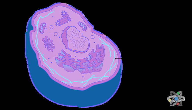
The important structural unit of the cytoskeleton is really a network of protein fibers in the cytoplasm that gives the cell its shape and enables it to perform important functions, such as cell movement.
Why Do Cells Need a Cytoskeleton?
Why Do Cells Need a Cytoskeleton?
While some people might imagine cells as unstructured, powerful microscopes used in cell biology reveal that cells are very organized.
One main component is vital to maintaining this shape and level of organization: the cytoskeleton of the cell. The protein filaments that make up the cytoskeleton form a network of fibers through the cell.
This network gives structural support to the plasma membrane, helps stabilize the organelles in their proper positions and enables the cell to shuffle its contents around as needed. For some cell types, the cytoskeleton even makes it possible for the cell to move and travel using specialized structures.
These form from the protein filaments when needed for cell locomotion.
The service the cytoskeleton provides for shaping the cell makes a lot of sense. Much like the human skeleton, the cytoskeleton protein network creates structural support that is crucial for maintaining the integrity of the cell and for preventing it from collapsing into its neighbors.
For cells with very fluid membranes, the network of proteins that make up the cytoskeleton are particularly important for keeping the cell contents inside the cell.
This is called membrane integrity.
Cytoskeleton Benefits for Cells
Cytoskeleton Benefits for Cells
Some highly specialized cells also rely on the cytoskeleton for structural support.
For these cells, maintaining the cell's unique shape makes it possible for the cell to function properly. These include neurons, or brain cells, which have round cell bodies, branchy arms called dendrites and stretched-out tails.
This characteristic cell shape makes it possible for neurons to catch signals using their dendrite arms and pass those signals through their axon tails and into the waiting dendrites of a neighboring brain cell. This is how brain cells communicate with each other.
It also makes sense that cells benefit from the organization that the cytoskeleton's protein fiber network gives them. There are over 200 types of cells in the human body and a grand total of about 30 trillion cells in each and every human on the planet.
The organelles in all these cells must perform a wide variety of cell processes, such as building and breaking down biomolecules, releasing energy for the body to use and performing a host of chemical reactions that make life possible.
For these functions to work well at a whole organism level, each cell needs a similar structure and way of doing things.
What Components Make up the Cytoskeleton
What Components Make up the Cytoskeleton
To perform those important roles, the cytoskeleton relies on three distinct types of filaments:
1. Microtubules 2. Intermediate filaments 3. Microfilaments
These fibers are all so infinitesimally small that they are completely invisible to the naked eye. Scientists only discovered them after the invention of the electron microscope brought the interior of the cell into view.
To visualize just how small these protein fibers are, it's helpful to understand the concept of the nanometer, which is sometimes written as nm. Nanometers are units of measurement just like an inch is a unit of measurement.
You might have guessed from the root word meter that the nanometer unit belongs to the metric system, just like a centimeter does.
Size Matters
Size Matters
Scientists use nanometers to measure extremely small things, such as atoms and light waves.
This is because one nanometer equals one billionth of a meter. This means that if you took a meter measuring stick, which is approximately 3 feet long when converted to the American system of measurement, and break it into one billion equal pieces, one single piece would equal one nanometer.
Now imagine that you could cut the protein filaments making up the cell's cytoskeleton and measure the diameter across the cut face.
Each fiber would measure between 3 and 25 nanometers in diameter, depending on the type of filament. For context, a human hair is 75,000 nanometers in diameter. As you can see, the filaments that make up the cytoskeleton are incredibly small.
Microtubules are the largest of the three fibers of the cytoskeleton, clocking in at 20 to 25 nanometers in diameter. Intermediate filaments are the cytoskeleton's mid-size fibers and measure about 10 nanometers in diameter.
The smallest protein filaments found in the cytoskeleton are microfilaments. These thread-like fibers measure a mere 3 to 6 nanometers in diameter.
In real-world terms, that is as much as 25,000 times smaller than the diameter of an average human hair.
Role of Microtubules in the Cytoskeleton
Role of Microtubules in the Cytoskeleton
Microtubules get their name from both their general shape and the type of protein they contain. They are tube-like and formed from repeating units of alpha- and beta-tubulin protein polymers linking together.
Read more about the main function of microtubules in cells.
If you were to view microtubule filaments under an electron microscope, they would look like chains of small proteins twisted together into a tight spiral lattice.
Each protein unit binds with all the units around it, producing a very strong, very rigid structure. In fact, microtubules are the most rigid structural component you can find in animal cells, which do not have cell walls like plant cells do.
But microtubules are not just rigid. They also resist compression and twisting forces. This quality increases the ability of the microtubule to maintain cell shape and integrity, even under pressure.
Microtubules also give the cell polarity, which means the cell has two unique sides, or poles. This polarity is part of what makes it possible for the cell to organize its components, such as organelles and other portions of the cytoskeleton, because it gives the cell a way to orient those components in relation to the poles.
Microtubules and Movement Within the Cell
Microtubules and Movement Within the Cell
Microtubules also support the movement of cell contents within the cell.
The microtubule filaments form tracks, which act like railroad tracks or highways in the cell. Vesicle transporters follow these tracks to move cell cargo around in the cytoplasm. These tracks are crucial for removing unwanted cell contents like misfolded proteins, old or broken organelles and pathogen invaders, such as bacteria and viruses.
Vesicle transporters simply follow the correct microtubule track to move this cargo to the cell's recycling center, the lysosome. There, the lysosome salvages and reuses some parts and degrades other parts.
The track system also helps the cell move newly built biomolecules, like proteins and lipids, out of the manufacturing organelles and to the places the cell needs the molecules.
For example, vesicle transporters use microtubule tracks to move cell membrane proteins from the organelles to the cell membrane.
Microtubules and Cell Movement
Microtubules and Cell Movement
Only some cells can use cell locomotion to travel, and those that do generally rely on specialized motile structures made of microtubule fibers.
The sperm cell is probably the easiest way to visualize these traveling cells.
As you know, sperm cells look a bit like tadpoles with long tails, or flagella, which they whip in order to swim to their destination and fertilize an egg cell. The sperm tail is made of tubulin and is an example of a microtubule filament used for cell locomotion.
Another well-known motile structure also plays a role in reproduction is the cilia. These hairlike motile structures line the fallopian tubes and use a waving motion to move the egg through the fallopian tube and into the uterus. These cilia are microtubule fibers.
Role of Intermediate Filaments in the Cytoskeleton
Role of Intermediate Filaments in the Cytoskeleton
Intermediate filaments are the second type of fiber found in the cytoskeleton. You can picture these as the true skeleton of the cell since their only role is structural support. These protein fibers contain keratin, which is a common protein you may recognize from body care products.
This protein makes up human hair and fingernails as well as the top layer of the skin. It is also the protein that forms horns, claws and hooves of other animals. Keratin is very strong and useful for protecting against damage.
The major role of intermediate filaments is the formation of the matrix of structural proteins under the cell membrane. This is like a supportive mesh that gives structure and shape to the cell. It also lends some elasticity to the cell, enabling it to respond flexibly under stress.
Intermediate Filaments and Organelle Anchoring
Intermediate Filaments and Organelle Anchoring
One of the important jobs performed by intermediate filaments is to help hold organelles in the right places within the cell. For example, intermediate filaments anchor the nucleus in its proper place within the cell.
This anchoring is crucial for cell processes because the various organelles inside a cell must work together to perform those cell functions. In the case of the nucleus, tethering this important organelle to the cytoskeleton matrix means that the organelles that rely on DNA instructions from the nucleus to do their jobs can easily access that information using messengers and transporters.
This important task might be impossible if the nucleus weren't anchored because those messengers and transporters would need to travel around searching through the cytoplasm for a wandering nucleus!
Role of Microfilaments in the Cytoskeleton
Role of Microfilaments in the Cytoskeleton
Microfilaments, also called actin filaments, are chains of actin proteins twisted into a spiral rod. This protein is best known for its role in muscle cells. There, they work with another protein called myosin to enable muscle contraction.
When it comes to the cytoskeleton, microfilaments aren't just the smallest fibers. They are also the most dynamic. Like all cytoskeleton fibers, microfilaments give the cell structural support. Because of their unique traits, microfilaments tend to show up at the edges of the cell.
The dynamic nature of actin filaments means that these protein fibers can change their lengths quickly to meet the cell's changing structural needs. This makes it possible for the cell to alter its shape or size or even form special projections that extend outside the cell, such as filopodia, lamellipodia and microvilli.
Microfilament Projections
Microfilament Projections
You can imagine filopodia as feelers that a cell projects to sense the environment around it, pick up chemical cues and even change the cell's direction, if it is moving. Scientists also sometimes call filopodia microspikes.
Filopodia can form part of another type of special projection, lamellipodia. This is a footlike structure that helps the cell move and travel.
Microvilli are like tiny hairs or fingers used by the cell during diffusion. The shape of these projections increases the surface area so that there is more space for molecules to move across the membrane through processes like absorption.
These fingers also perform a fascinating function called cytoplasm streaming.
This occurs when the actin filaments comb through the cytoplasm to keep it moving. Cytoplasm streaming boosts diffusion and helps moves wanted materials, like nutrients, and unwanted materials, like waste and cell debris, around in the cell.
Cite This Article
MLA
Mayer, Melissa. "Cytoskeleton: Definition, Structure & Function (With Diagram)" sciencing.com, https://www.sciencing.com/cytoskeleton-definition-structure-function-with-diagram-13717285/. 29 July 2019.
APA
Mayer, Melissa. (2019, July 29). Cytoskeleton: Definition, Structure & Function (With Diagram). sciencing.com. Retrieved from https://www.sciencing.com/cytoskeleton-definition-structure-function-with-diagram-13717285/
Chicago
Mayer, Melissa. Cytoskeleton: Definition, Structure & Function (With Diagram) last modified March 24, 2022. https://www.sciencing.com/cytoskeleton-definition-structure-function-with-diagram-13717285/

