Difference Between Compound & Dissecting Microscopes
Dissecting and compound light microscopes are both optical microscopes that use visible light to create an image. Both types of microscope magnify an object by focusing light through prisms and lenses, directing it toward a specimen, but differences between these microscopes are significant. Most importantly, dissecting microscopes are for viewing the surface features of a specimen, whereas compound microscopes are designed to look through a specimen.
How a Microscope Works
How a Microscope Works
Both dissecting and compound light microscopes work by capturing and redirecting light reflected and refracted from a specimen. Compound microscopes also capture light that is transmitted through a specimen. Light is captured by bi-convex lenses above the specimen; these are called objective lenses. Compound microscopes have several objective lenses of varying strengths, magnifying from 40 to 1,000 times. The point at which the light is redirected — or converged — is called the focal point. The image at the focal point will appear magnified to the observer. The distance between the focal point and the first lens is called the working distance. Microscopes with a smaller working distance have greater magnifying power than those with a longer one.
Dissecting Microscopes
Dissecting Microscopes
The dissecting microscope is also known as a stereomicroscope. Because it has a long working distance, between 25 and 150 mm, it has a lower magnification ability. This gives the user the option to manipulate the specimen, even performing small dissections under the microscope. Live specimens can also be observed. A typical student stereoscope can magnify two to 70 times through its one objective lens. With a stereoscope, light can be directed at the specimen from above, creating a three dimensional image.
Compound Microscopes
Compound Microscopes
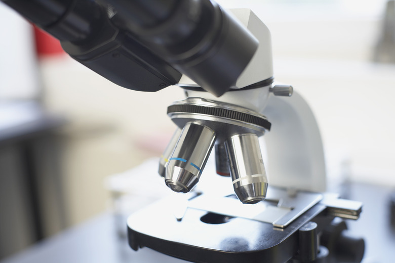
Compound light microscopes are commonly used to view items that are too small to see with the naked eye. They have several strengths of objective lenses and rely on light shining from beneath the specimen. This requires that a specimen be very thin and at least partly translucent. Most specimens are stained, sectioned and placed on a glass slide for viewing. A compound microscope can magnify up to 1,000 times and provide the ability to see much more detail. The working distance varies from 0.14 to 4 mm.
Differences in Application
Differences in Application
A compound microscope is used to observe ultra-thin pieces of larger objects. Examples could be the stem of a plant or a cross section of a human blood vessel. In both cases, the specimen is not living. The piece is placed on a slide and stained with dyes to highlight features. A stereoscope can be used for items that light cannot shine through. The actual colors of the specimen will be observed, and the specimen can be manipulated by the observer while being viewed. The intricacy of butterfly wings, the detail of a scorpion claw and the weave in a fabric are a few examples of items that could be viewed. Stereoscopes also might be used to observe some living organisms such as those in pond water.
Cite This Article
MLA
Taylor, Stacy. "Difference Between Compound & Dissecting Microscopes" sciencing.com, https://www.sciencing.com/difference-between-compound-dissecting-microscopes-5576645/. 9 March 2018.
APA
Taylor, Stacy. (2018, March 9). Difference Between Compound & Dissecting Microscopes. sciencing.com. Retrieved from https://www.sciencing.com/difference-between-compound-dissecting-microscopes-5576645/
Chicago
Taylor, Stacy. Difference Between Compound & Dissecting Microscopes last modified March 24, 2022. https://www.sciencing.com/difference-between-compound-dissecting-microscopes-5576645/
