Difference Between Working Distance & Magnification
Microscopes have been around in some form for centuries. The human urge to explore what the unaided eye alone cannot has led to innovations such as telescopes, microscopes and infrared ("night vision") optical equipment, and the storehouse of science knowledge to which you now have access is a remarkable reward.
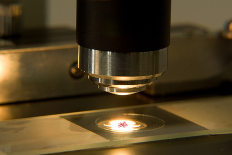
The primary job of a microscope is to produce magnification of a specimen or other object. This means interposing a number of instruments between the specimen to be examined and your own eyes, chiefly lenses (usually more than one). Also important is the distance between the specimen and the first lens the light waves reflected from the specimen encounter, known as the working distance.
Parts of a Compound Microscope
Parts of a Compound Microscope
This discussion describes light microscopes, as most modern microscopes are equipped with their own light source. The specimen usually sits in a translucent (light-transmitting) side on a level stage. The distance between the stage and the objective lens is controlled by a rotating knob that allows exquisite focus of a properly prepared specimen.
Compound microscopes get their name from the fact that they have two lens systems. The objective lens system normally has multiple magnification options, each of which is positioned over the specimen by rotating a dial, while the other system is called the eyepiece or optical lens. Usually there is only one of these.
It is also possible to control the amount of light passing upward through the specimen area by using the diaphragm, which makes the circular opening transmitting light larger or smaller.
Magnification Explained
Magnification Explained
The objective lens system has individually labeled lenses, often 10x, 40x and 100x. The eyepiece lens is typically 10x. Magnification is simply making an object appear larger by in effect reducing the distance between your eyes and the specimen to a degree that would be impossible without the systematic manipulation of light waves.
**The total magnification for a given viewing is found by multiplying the objective lens magnification by the eyepiece magnification.** For example, a specimen viewed using a combination of 40x and 10x would appear 400 times larger than it would if you looked at it from the same distance using your eyes alone; clearly, this can be the difference between seeing something in considerable detail and not being able to see even a tiny dot.
Working Distance of a Microscope
Working Distance of a Microscope
The working distance of a microscope, defined as the distance between the objective lens and the specimen, is controlled by moving the stage up and down. There are usually two knobs for this, one that moves the stage up and down in tiny increments (fine focus) and another than moves it in greater increments (coarse focus).
When you first use a microscope, it's a good idea to experiment with the controls other than the lenses, which can quickly captivate your attention thanks to the wonders they often reveal. In particular, try to avoid pressing the objective lens into the specimen itself and damaging or destroying it.
Relationship Between Magnification and Working Distance
Relationship Between Magnification and Working Distance
**Working distance and magnification are inversely related.** This means that as you increase the magnification, you have to move the lens closer to the specimen to achieve an optimal image.
Thus at lower levels of magnification, the ideal working distance is comparatively long. As you increase the magnification, the working distance decreases very rapidly. Oil-immersion lenses, which are often used for 100x objective lenses, are very, very close the specimen when optimal focus is achieved. As noted above, this can lead to accidental damage to the specimen and a possible compromise of your work. So, be patient while enjoying the benefits of an astonishing yet simple piece of science equipment!
References
Cite This Article
MLA
Beck, Kevin. "Difference Between Working Distance & Magnification" sciencing.com, https://www.sciencing.com/difference-between-working-distance-magnification-7203011/. 9 November 2019.
APA
Beck, Kevin. (2019, November 9). Difference Between Working Distance & Magnification. sciencing.com. Retrieved from https://www.sciencing.com/difference-between-working-distance-magnification-7203011/
Chicago
Beck, Kevin. Difference Between Working Distance & Magnification last modified March 24, 2022. https://www.sciencing.com/difference-between-working-distance-magnification-7203011/
