Do Prokaryotes Have Cell Walls?

Prokaryotes represent one of the two major classifications of life. The others are the eukaryotes.
Prokaryotes are set apart by their lower level of complexity. They are all microscopic, though not necessarily unicellular. They are divided into the domains archaea and bacteria, but the vast majority of known prokaryote species are bacteria, which have been on Earth for around 3.5 billion years.
Prokaryotic cells do not have nuclei or membrane-bound organelles. 90 percent of bacteria do, however, have cell walls, which, with the exception of plant cells and some fungal cells, eukaryotic cells lack. These cell walls form the outermost layer of bacteria and make up part of the bacterial capsule.
They stabilize and protect the cell and are vital to bacteria being able to infect host cells as well as the bacteria's response to antibiotics.
General Characteristics of Cells
General Characteristics of Cells
All cells in nature share many features in common. One of these is the presence of an external _cell membrane_, or plasma membrane, which forms the physical boundary of the cell on all sides. Another is the substance known as cytoplasm found within the cell membrane.
A third is the inclusion of genetic material in the form of DNA, or deoxyribonucleic acid. A fourth is the presence of ribosomes, which manufacture proteins. Every living cell uses ATP (adenosine triphosphate) for energy.
General Prokaryotic Cell Structure
General Prokaryotic Cell Structure
The structure of prokaryotes is simple. In these cells, DNA, rather than being packaged within a nucleus enclosed within a nuclear membrane, is found more loosely gathered in the cytoplasm, in the form of a body called a nucleoid.
This is normally in the form of a circular chromosome.
The ribosomes of the prokaryotic cell are found scattered throughout the cell cytoplasm, whereas in eukaryotes, some of them are found in organelles such as the Golgi apparatus and the endoplasmic reticulum. Ribosomes' job is protein synthesis.
Bacteria reproduce by binary fission, or simply splitting in two and dividing the cell components equally, including the genetic information in the single small chromosome.
Unlike mitosis, this form of cell division does not require distinct stages.
Structure of the Bacterial Cell Wall
Structure of the Bacterial Cell Wall
**The Unique Peptidoglycans:** All plant cell walls and bacterial cell walls consist mostly of carbohydrate chains.
But while plant cell walls contain cellulose, which you will see listed in the ingredients of numerous foods, the walls of bacterial cells contain a substance called peptidoglycan, which you will not.
This peptidoglycan, which is found only in prokaryotes, comes in different types; it gives the cell as a whole its shape and confers protection for the cell from mechanical insults.
Peptidoglycans consist of a backbone called glycan, which itself consists of muramic acid and glucosamine, both of which in turn have acetyl groups attached to their nitrogen atoms. They also include peptide chains of amino acids that are cross-linked to other, nearby peptide chains.
The strength of these "bridging" interactions varies widely between different peptidoglycans and therefore between different bacteria.
This characteristic, as you'll see, allows bacteria to be classified into distinct types based on how their cell walls react to a certain chemical.
The cross-links are formed by the action of an enzyme called a transpeptidase, which is the target of a class of antibiotics used to combat infectious disease in humans and other organisms.
Gram-Positive and Gram-Negative Bacteria
Gram-Positive and Gram-Negative Bacteria
While all bacteria have a cell wall, its composition changes from species to species owing to differences in the peptidoglycan content of which the cell walls are partly or mostly made.
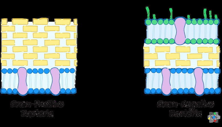
Bacteria may be separated into two types called gram-positive and gram-negative.
These are named after the biologist Hans Christian Gram, a pioneer in cell biology who developed a staining technique in the 1880s, aptly called the Gram stain, that caused certain bacteria to become purple or blue and others to become red or pink.
The former type of bacteria came to be known as gram-positive, and their staining properties are attributable to the fact that their cell walls contain a very high fraction of peptidoglycan in relation to the entirety of the wall.
The red or pink-staining bacteria are known as gram-negative, and as you might guess, these bacteria have walls that consist of modest to small amounts of peptidoglycan.
In gram-negative bacteria, a thin membrane lies outside the cell wall, forming the cell envelope.
This layer is similar to the plasma membrane of the cell that lies on the other side of the cell wall, closer to the interior of the cell. In some gram-negative cells, such as E. coli, the cell membrane and the nuclear envelope actually come into contact in some places, penetrating the peptidoglycan of the thin wall between.
This nuclear envelope contains outward-extending molecules called **lipopolysaccharides,** or LPS. Extending from the interior of this membrane are murein lipoproteins that are attached at the far end to the outside of the cell wall.
Gram-Positive Bacterial Cell Walls
Gram-Positive Bacterial Cell Walls
Gram-positive bacteria have a thick peptidoglycan cell wall, about 20 to 80 nm (nanometers or one-billionths of a meter) thick.
Examples include staphylococci, streptococci, lactobacilli and Bacillus species.
These bacteria stain purple or red, but usually purple, with Gram stain, as the peptidoglycan retains the violet dye applied early in the procedure when the preparation is later washed with alcohol.
This more robust cell wall offers gram-positive bacteria more protection from most outside insults compared to gram-negative bacteria, although the high peptidoglycan content of these organisms makes their walls something of a one-dimensional fortress, making in turn for a somewhat easier strategy concerning how to destroy it.
 Sciencing
Gram-positive bacteria are generally more susceptible to antibiotics that target the cell wall than are gram-negative species, since it is exposed to the environment as opposed to sitting beneath, or within, a cell envelope.
The Role of Teichoic Acids
The Role of Teichoic Acids
The peptidoglycan layers of gram-positive bacteria are usually high in molecules called teichoic acids, or TAs.
These are carbohydrate chains that reach through and sometimes past the peptidoglycan layer.
TA is believed to stabilize the peptidoglycan around it simply by making it more rigid, rather than by exerting any chemical properties.
TA is in part responsible for the ability of certain gram-positive bacteria, such as Streptococcal species, to bind to specific proteins on the surface of host cells, which facilitates their ability to cause infection and in many cases disease.
When bacteria or other microorganisms are capable of causing infectious disease, they are referred to as pathogenic.
The cell walls of bacteria of the Mycobacteria family, in addition to containing peptidoglycan and TAs, have an external "waxy" layer of made of mycolic acids. These bacteria are known as "acid-fast," because stains of this type are needed to penetrate this waxy layer to permit useful microscopic examination.
Gram-Negative Bacterial Cell Walls
Gram-Negative Bacterial Cell Walls
Gram-negative bacteria, like their gram-positive counterparts, have peptidoglycan cell walls.
However, the wall is much thinner, only about 5 to 10 nm thick. These walls do not stain purple with Gram stain because their smaller peptidoglycan content means the wall cannot retain much dye when the preparation is washed with alcohol, resulting in a pink or reddish color in the end.
As noted above, the cell wall is not the outermost later of these bacteria but is instead covered by another plasma membrane, the cell envelope or outer membrane.
This layer is about 7.5 to 10 nm thick, rivaling or exceeding the thickness of the cell wall.
In most gram-negative bacteria, the cell envelope is linked to a type of lipoprotein molecule called Braun's lipoprotein, which, in turn is linked to the peptidoglycan of the cell wall.
The Tools of Gram-Negative Bacteria
The Tools of Gram-Negative Bacteria
Gram-negative bacteria are generally less susceptible to antibiotics targeting the cell wall because it is not exposed to the environment; it still has the outer membrane for protection.
In addition, in gram-negative bacteria, a gel-like matrix occupies the territory inside the cell wall and outside the plasma membrane called the periplasmic space.
The peptidoglycan component of the cell wall of gram-negative bacteria is only about 4 nm thick.
Where a gram-positive bacterial cell wall would have more peptidoglycans to give its wall substance, a gram-negative bug has other tools in store in its outer membrane.
Each LPS molecule is composed of a fatty acid-rich Lipid A subunit, a small core polysaccharide and an O-side chain made of sugar-like molecules. This O-side chain forms the external side of the LPS.
The exact composition of the side chain varies between different bacterial species.
Portions of the O-side chain known as antigens can be identified via laboratory tests to identify specific pathogenic bacterial strains (a "strain" is a subtype of a bacterial species, like a breed of dog).
Archaea Cell Walls
Archaea Cell Walls
Archaea are more diverse than bacteria and so are their cell walls. Notably, these walls do not contain peptidoglycan.
Rather, they usually contain a similarly called molecule called pseudopeptidoglycan, or pseudomurein. In this substance, a portion of regular peptidoglycan called NAM is replaced with a different subunit.
Some archaea may instead have a layer of glycoproteins or polysaccharides that substitute for the cell wall in place of pseudopeptidoglycan. Finally, as with some bacterial species, a few archaea are missing cell walls altogether.
Archaea that contain pseudomurein are insensitive to antibiotics of the penicillin class because these drugs are transpeptidase inhibitors that act to interfere with peptidoglycan synthesis.
In these archaea, there are no peptidoglycans being synthesized and therefore nothing for penicillins to act on.
Why Is the Cell Wall Important?
Why Is the Cell Wall Important?
Bacterial cells lacking cell walls may have additional cell surface structures in addition to those discussed, such as glycocalyces (singular is glycocalyx) and S-layers.
A glycocalyx is a coat of sugar-like molecules that comes in two main types: capsules and slime layers. A capsule is a well-organized layer of polysaccharides or proteins. A slime layer is less tightly organized, and it is less tightly attached to the cell wall below than a glycocalyx.
As a result, a glycocalyx is more resistant to being washed away, while a slime layer can be more easily displaced. The slime layer may be composed of polysaccharides, glycoproteins or glycolipids.
These anatomical variations lend themselves to great clinical significance.
Glycocalyces allow cells to stick to certain surfaces, assisting in the formation of colonies of organisms called biofilms that can form several layers and protect the individuals in the group. For this reason, most bacteria in the wild live in biofilms formed from mixed bacterial communities. Biofilms impede the action of antibiotics as well as disinfectants.
All of these attributes contribute to the difficulty of eliminating or reducing microbes and eradicating infections.
Antibiotic Resistance
Antibiotic Resistance
Bacterial strains that are naturally resistant to a given antibiotic thanks to a chance advantageous mutation are "selected for" in human populations because these are the bugs left behind when the antibiotic-susceptible ones are killed off, and these "superbugs" multiply and continue to cause disease.
By the second decade of the 21st century, a variety of gram-negative bacteria has become increasingly resistant to antibiotics, leading to increased illness and death from infections and driving up health care costs. Antibiotic resistance is an archetypal example of natural section on time scales observable to humans.
Examples include:
- E. coli, which causes urinary-tract infections (UTIs).
- Acinetobacter baumanii, which causes problems mainly in health care settings.
- Pseudomonas aeruginosa, which causes blood infections and pneumonia in hospitalized patients and pneumonia in patients with the inherited disease cystic fibrosis.
- Klebsiella pneumoniae, which is responsible for a lot of infections in health care-associated settings, among them pneumonia, blood infections and UTIs.
- Neisseria gonorrhoeae, which causes the sexually transmitted disease gonorrhea, the second most commonly reported infectious disease in the U.S.
Medical researchers are working to keep up with resistant bugs in what amounts to a microbiological arms race.
Cite This Article
MLA
Beck, Kevin. "Do Prokaryotes Have Cell Walls?" sciencing.com, https://www.sciencing.com/do-prokaryotes-have-cell-walls-13717681/. 31 July 2019.
APA
Beck, Kevin. (2019, July 31). Do Prokaryotes Have Cell Walls?. sciencing.com. Retrieved from https://www.sciencing.com/do-prokaryotes-have-cell-walls-13717681/
Chicago
Beck, Kevin. Do Prokaryotes Have Cell Walls? last modified March 24, 2022. https://www.sciencing.com/do-prokaryotes-have-cell-walls-13717681/