What Does A Human Cell Look Like?
Exploring the human cell quickly reveals to us a complex structure made of many parts. It can seem impossible that something so tiny is made up of so many moving parts, but inside a human cell, there are several unique shapes, textures and sizes.
All play critical roles in making sure that each cell does its job efficiently. Some of those shapes may look familiar, while others may be new to you.
Cell Membrane

Cell Membrane
To imagine the cell membrane, think of a swimming pool. View the pool as having tennis balls covering both the surface and the bottom of the pool. The cell membrane consists of the tennis balls on the surface and bottom of the pool. The membrane works to protect the cell, and also is responsible for letting substances into the cell and its organelles.
In humans and animals, the cell membrane protects the cell's contents in a roughly circular shape. In plants, though, the membrane and wall are shaped like a rectangular box.
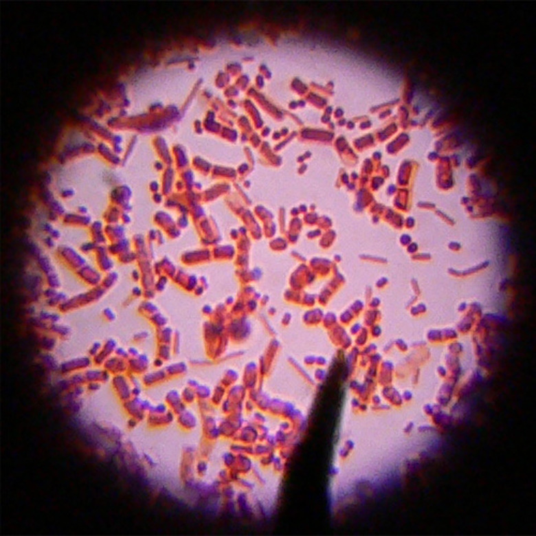
Rough Endoplasmic Reticulum
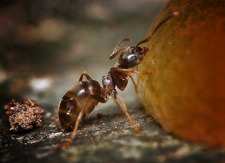
Rough Endoplasmic Reticulum
The RER (rough endoplasmic reticulum) looks similar to an ant farm where all the ants travel along a wavy and uniform path. Space each ant an even distance from the ant in front and the ant behind it. This description is more accurate if we string the ants together using a thread. Now replace each ant's body with a circular dot and that's what the RER looks like.
Smooth Endoplasmic Reticulum
Smooth Endoplasmic Reticulum
Using the previous description of what RER looks like, we can draw a quick basis for what SER (smooth endoplasmic reticulum) looks like. Imagine the same ant farm with its wavy tunnels and simply remove the ants. SER will look similar with the primary difference being that the paths are smoother in shape.
Nucleus and Nucleolus

Nucleus and Nucleolus
The nucleus is an important part of the cell, and in an animal cell, it's also the largest organelle. The nucleus has a body that looks similar in shape to an orange. Using your imagination, cover the surface of the nucleus with dips similar to the surface of a strawberry. These two fruits will aid in remembering the unique shape and surface texture of a nucleus.
Placed within the nucleus is the nucleolus. Looking at the nucleolus may remind you of the pit of an avocado. Placing the pit of an avocado inside the center of the nucleus will give you a fair representation of the shape, size and location of the nucleolus.
Mitochondria and Lysosome

Mitochondria and Lysosome
The shape of a mitochondrion is oval, similar to a pharmaceutical capsule. The inside looks much like a winding river which meanders back and forth. The winding river may have occasional islands along its path.
Use the shape of a basketball to suggest the external appearance of a lysosome. Now place several small grapes inside this basketball to represent the active hydrolytic enzymes contained within a lysosome.
Ribosome

Ribosome
The ribosome is one of the most complex parts of the inside of a cell. A large mass of silly string floating in the air offers the mind a vivid image of what a ribosome looks like. Add some long spiral-shaped confetti and the image of what a ribosome looks like will be even more accurate.
A Quick Wrap-Up

A Quick Wrap-Up
No description can compare with a firsthand observation of a human cell through a powerful microscope. If you have a chance to do so, you'll be able to see how many different shapes and textures are present in every single cell.
Incredibly, the human body has trillions of those, all working to help keep our bodies doing all the things they need to do. The descriptions above will help paint a general picture of a human cell looks like in terms that are visually pleasing and easy to remember.
Cite This Article
MLA
Stokes, Aaron L.. "What Does A Human Cell Look Like?" sciencing.com, https://www.sciencing.com/human-cell-look-like-5614496/. 25 March 2019.
APA
Stokes, Aaron L.. (2019, March 25). What Does A Human Cell Look Like?. sciencing.com. Retrieved from https://www.sciencing.com/human-cell-look-like-5614496/
Chicago
Stokes, Aaron L.. What Does A Human Cell Look Like? last modified March 24, 2022. https://www.sciencing.com/human-cell-look-like-5614496/