Mitochondria: Definition, Structure & Function (With Diagram)
The eukaryotic cells of living organisms continuously carry out a huge number of chemical reactions to live, grow, reproduce and fight off disease.
All these processes require energy at the cellular level. Each cell that engages in any of these activities gets its energy from the mitochondria, tiny organelles that act as the cells' powerhouses. The singular of mitochondria is mitochondrion.
In humans, cells such as red blood corpuscles don't have these tiny organelles, but most other cells have large numbers of mitochondria. Muscle cells, for example, may have hundreds or even thousands to satisfy their energy requirements.
Almost every living thing that moves, grows or thinks has mitochondria in the background, producing the necessary chemical energy.
Structure of the Mitochondria
Structure of the Mitochondria
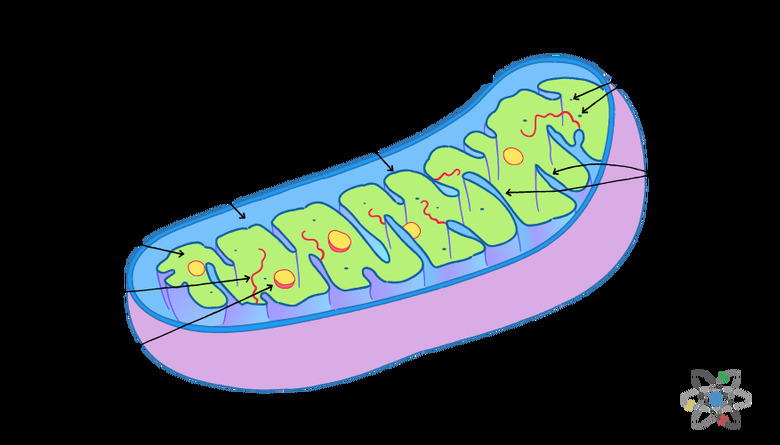
Mitochondria are membrane-bound organelles enclosed by a double membrane.
They have a smooth outer membrane enclosing the organelle and a folded inner membrane. The folds of the inner membrane are called cristae, the singular of which is crista, and the folds are where the reactions creating mitochondrial energy take place.
The inner membrane contains a fluid called the matrix while the intermembrane space located between the two membranes is also filled with fluid.
Because of this relatively simple cell structure, mitochondria have only two separate operating volumes: the matrix inside the inner membrane and the intermembrane space. They rely on transfers between the two volumes for energy generation.
To increase efficiency and maximize energy creation potential, the inner membrane folds penetrate deep into the matrix.
As a result, the inner membrane has a large surface area, and no part of the matrix is far from an inner membrane fold. The folds and large surface area help with the mitochondrial function, increasing the potential rate of transfer between the matrix and the intermembrane space across the inner membrane.
Why Are Mitochondria Important?
Why Are Mitochondria Important?
While single cells originally evolved without mitochondria or other membrane-bound organelles, complex multicellular organisms and warm-blooded animals such as mammals get their energy from cellular respiration based on the mitochondrial function.
High-energy functions such as those of the heart muscles or bird wings have high concentrations of mitochondria that supply the energy needed.
Through their ATP synthesis function, mitochondria in muscles and other cells produce the body heat to keep warm-blooded animals at a steady temperature. It is this concentrated energy production capability of mitochondria that makes the high-energy activities and the production of heat in higher animals possible.
Mitochondrial Functions
Mitochondrial Functions
The energy-production cycle in mitochondria relies on the an electron transport chain along with the citric acid or Krebs cycle.
Read more about the Krebs Cycle.
The process of breaking down carbohydrates such as glucose to make ATP is called catabolism. The electrons from glucose oxidation are passed along a chemical reaction chain that includes the citric acid cycle.
Energy from the reduction-oxidation, or redox, reactions is used to transfer protons out of the matrix where the reactions are taking place. The final reaction in the mitochondrial function chain is one in which oxygen from cellular respiration undergoes reduction to form water. The end products of the reactions are water and ATP.
The key enzymes responsible for mitochondrial energy production are nicotinamide adenine dinucleotide phosphate (NADP), nicotinamide adenine dinucleotide (NAD), adenosine diphosphate (ADP) and flavin adenine dinucleotide (FAD).
They work together to help transfer protons from hydrogen molecules in the matrix across the inner mitochondrial membrane. This creates a chemical and electrical potential across the membrane with the protons returning to the matrix through the enzyme ATP synthase, resulting in the phosphorylation and production of adenosine triphosphate (ATP).
Read about the structure and function of ATP.
ATP synthesis and the ATP molecules are the prime carriers of energy in cells and can be used by the cells for the production of the chemicals necessary for living organisms.
In addition to being energy producers, mitochondria can help with cell-to-cell signaling through the release of calcium.
Mitochondria have the ability to store calcium in the matrix and can release it when certain enzymes or hormones are present. As a result, cells producing such triggering chemicals may see the signal of rising calcium from the release by the mitochondria.
Overall, mitochondria are a vital component of living cells, helping with cell interactions, distributing complex chemicals and producing the ATP that forms the energy basis for all life.
The Inner and Outer Mitochondrial Membranes
The Inner and Outer Mitochondrial Membranes
The mitochondrial double membrane has different functions for the inner and outer membrane and the two membranes and are made up of different substances.
The outer mitochondrial membrane encloses the fluid of the intermembrane space, but it has to allow chemicals that the mitochondria need to pass through it. Energy-storage molecules produced by the mitochondria have to be able to leave the organelle and deliver energy to the rest of the cell.
To allow for such transfers, the outer membrane is made up of phospholipids and protein structures called porins that leave tiny holes or pores in the surface of the membrane.
The intermembrane space contains fluid that has a composition similar to that of the cytosol making up the fluid of the surrounding cell.
Small molecules, ions, nutrients and the energy-carrying ATP molecule produced by ATP synthesis can penetrate the outer membrane and transition between the fluid of the intermembrane space and the cytosol..
The inner membrane has a complex structure with enzymes, proteins and fats allowing only water, carbon dioxide and oxygen to pass through the membrane freely.
Other molecules, including large proteins, can penetrate the membrane but only through special transport proteins that limit their passage. The large surface area of the inner membrane, resulting from the cristae folds, provides room for all these complex protein and chemical structures.
Their large number permits a high level of chemical activity and an efficient production of energy.
The process by which energy is produced through chemical transfers across the inner membrane is called oxidative phosphorylation.
During this process, the oxidation of carbohydrates in the mitochondria pumps protons across the inner membrane from the matrix into the intermembrane space. The imbalance in protons causes the protons to diffuse back across the inner membrane into the matrix through an enzyme complex that is a precursor form of ATP and is called ATP synthase.
The flow of protons through ATP synthase in turn is the basis for ATP synthesis and it produces ATP molecules, the main energy-storage mechanism in cells.
What’s in the Matrix?
What's in the Matrix?
The viscous fluid inside the inner membrane is called the matrix.
It interacts with the inner membrane to carry out the main energy-producing functions of the mitochondria. It contains the enzymes and chemicals that take part in the krebs cycle to produce ATP from glucose and fatty acids.
The matrix is where the mitochondrial genome made up of circular DNA is found and where the ribosomes are located. The presence of ribosomes and DNA means that the mitochondria can produce their own proteins and can reproduce using their own DNA, without relying on cell division.
If mitochondria seem to be tiny, complete cells on their own, it is because they were probably separate cells at one point when single cells were still evolving.
Mitochondrion-like bacteria entered larger cells as parasites and were allowed to remain because the arrangement was mutually beneficial.
The bacteria were able to reproduce in a secure environment and supplied energy to the larger cell. Over hundreds of millions of years, the bacteria became integrated into multicellular organisms and evolved into today's mitochondria.
Because they are found in animal cells today, they form a key part of early human evolution.
Since mitochondria multiply independently based on the mitochondrial genome and don't take part in cell division, new cells simply inherit the mitochondria that happen to be in their part of the cytosol when the cell divides.
This function is important for the reproduction of higher organisms, including humans, because embryos develop from a fertilized egg.
The egg cell from the mother is large and contains a lot of mitochondria in its cytosol while the fertilizing sperm cell from the father has hardly any. As a result, children inherit their mitochondria and their mitochondrial DNA from their mother.
Through their ATP synthesis function in the matrix and through cellular respiration across the double membrane, mitochondria and the mitochondrial function are a key component of animal cells and help make life as it exists possible.
Cell structure with membrane-bound organelles has played an important part in human evolution and mitochondria have made an essential contribution.
Cite This Article
MLA
Markgraf, Bert. "Mitochondria: Definition, Structure & Function (With Diagram)" sciencing.com, https://www.sciencing.com/mitochondria-definition-structure-function-with-diagram-13717287/. 30 July 2019.
APA
Markgraf, Bert. (2019, July 30). Mitochondria: Definition, Structure & Function (With Diagram). sciencing.com. Retrieved from https://www.sciencing.com/mitochondria-definition-structure-function-with-diagram-13717287/
Chicago
Markgraf, Bert. Mitochondria: Definition, Structure & Function (With Diagram) last modified August 30, 2022. https://www.sciencing.com/mitochondria-definition-structure-function-with-diagram-13717287/

