The Purpose Of Electrophoresis
Electrophoresis is a "powerful and inexpensive molecular separation technique," as stated by Dr. William H. Heidcamp, in the Cell Biology Laboratory Manual. Various reasons exist for carrying out electrophoresis including non-invasive binding to molecules and visualization of molecule separation. Overall, electrophoresis aims to provide an accurate way of analyzing substances, such as your blood and DNA (deoxyribonucleic acid), which are difficult to separate using conventional methods.
Definition
Definition
Electrophoresis is an empirical technique used in the separation of charged molecules (positive and negative) such as cells and proteins, according to their response in electric current.
Several factors affect electrophoresis, including net charge, mass of molecule, buffer and electrophoretic media like paper or gel. In electrophoresis, molecules move towards the opposite charge; for instance, a protein with a positive net charge move towards the negative side of the electrophoretic medium. Furthermore, molecules with smaller mass move faster or separate faster than molecules with larger mass.
History
History
In 1937, a Swedish scientist named Arne Tiselius developed an apparatus for measuring the movement of protein molecules, called Moving Boundary apparatus. This is a U-shaped apparatus that uses an aqueous medium for separating protein molecules.
In 1940, zone electrophoresis was introduced, which uses a solid medium (e.g., gel) and allows staining for better resolution or visualization of the separation of molecules.
Then in 1960, the capillary electrophoresis was developed to provide a versatile electrophoresis technique. This type of electrophoresis allows separation of molecules using aqueous and solid mediums.
Molecule Binding
Molecule Binding
Electrophoresis, using mediums, purposely interact with molecules in a non-invasive way. For instance, gel mediums bind to protein molecules without disrupting the protein's structure and function. After binding to molecules, movement or separation is initiated by applying electric current. Furthermore, it is also possible to recover the molecules bound to the medium after electrophoresis.
High-Resolution Separation
High-Resolution Separation
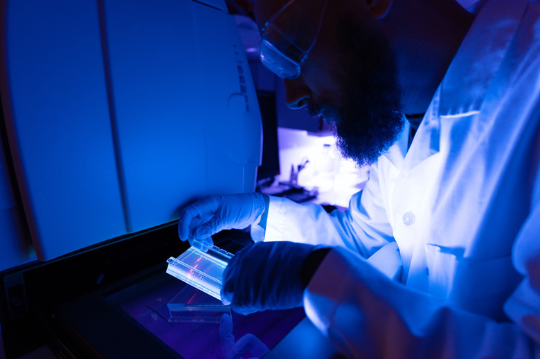
Electrophoresis is designed to visualize the separation of molecules. This is achieved by various methods, including staining and autoradiography.
Autoradiography uses X-ray films to visualize the position of radioactive molecules (e.g., DNA) after separation. This type of visualization is comparable to taking pictures, wherein the x-ray is like a camera flash and the x-ray film is like the film used in developing black-and-white photos. In electrophoresis, photos of molecules such as proteins in your blood are developed using autoradiography.
In staining, dyes such as coomassie blue and amido black are mixed with molecules, before or after the separation process. For instance, mixing proteins with coomasie dye prior to electrophoresis will yield stained paths (small dots or lines) showing the movement of protein during separation.
Quantitative Analysis
Quantitative Analysis
Another purpose of electrophoresis is to obtain quantitative information after visualizing the separation of molecules. To obtain quantitative data, for instance, image analysis software (2D and 3D rendering software) records the results of electrophoresis as digital signals. These signals represent the molecules' position before and after electrophoresis and are then used for quantitative analysis 'in silico' (with the use of a computer).
Cite This Article
MLA
Dephoff, Jacklyn. "The Purpose Of Electrophoresis" sciencing.com, https://www.sciencing.com/purpose-electrophoresis-5426937/. 18 September 2009.
APA
Dephoff, Jacklyn. (2009, September 18). The Purpose Of Electrophoresis. sciencing.com. Retrieved from https://www.sciencing.com/purpose-electrophoresis-5426937/
Chicago
Dephoff, Jacklyn. The Purpose Of Electrophoresis last modified August 30, 2022. https://www.sciencing.com/purpose-electrophoresis-5426937/
