The Reason For Staining A Specimen On The Microscope
The main reason you stain a specimen before putting it under the microscope is to get a better look at it, but staining does much more than simply highlight the outlines of cells. Some stains can penetrate cell walls and highlight cell components, and this can help scientists visualize metabolic processes. Stains also help distinguish between live cells and dead ones. Moreover, staining allows scientists count the number of cells of a particular type within a certain biomass. Twenty or more different types of stains exist, and each one has its purpose.
TL;DR (Too Long; Didn't Read)
The main purpose of staining is to highlight cells and parts of cells. Over 20 different types of stains exist, and the type of stain you use depends on what you are looking for.
Types of Stains
Types of Stains
The choice of stain depends what you are looking for. Not all stains are suitable for living cells, but those that are include Bismarck brown, toluene red, Nile blue and Nile red, and certain fluorescents used to highlight DNA. Some stains highlight spores, some detect lipids and proteins, and some change color in the presence of starches. The purpose of the examination determines the best type of stain to use. For example, a medical practitioner conducting a PAP smear would use Eosin Y. It's an acidic fluorescent dye that turns red when it contacts red blood cells, cytoplasm and cell membranes. It's also used to test blood marrow.
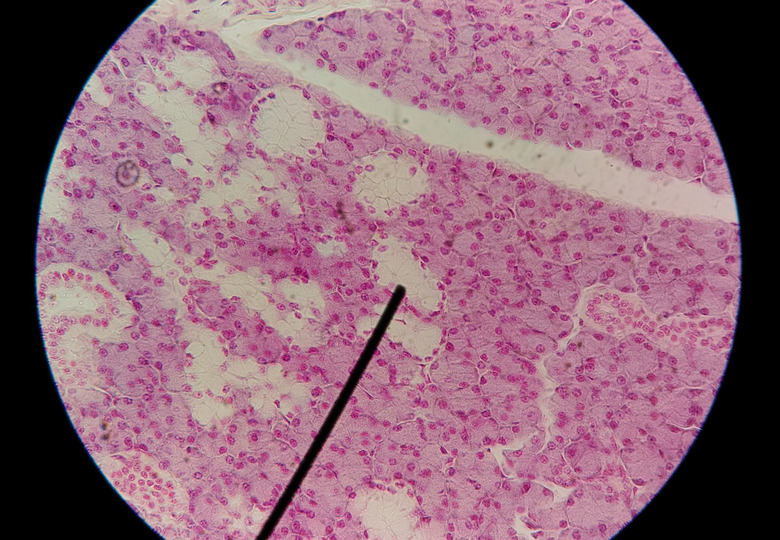
In some cases, an investigator may use more than one stain. For example, hematoxylin is a stain that turns cell nuclei blue. When used in conjunction with eosin, which turns the other parts of the cell red or pink, it provides a stronger contrast and makes the nuclei easier to differentiate. PAP smears and blood marrow samples are easier to examine when these two stains are used together.
**Gram's Stain:** Hospital workers use Gram's stain to identify harmful bacteria. This is actually a series of colorants that have different effects on different types of bacteria and give doctors an important diagnostic tool. Gram's stain is a three-part process. In the first, Hucker's crystal violet is added, which stains all bacteria a uniform violet color. In the next stage, iodine stain is added, which causes the color to adhere to Gram-positive cells, which are primarily Staphylococcus and Streptococcus. The stain is washed away, leaving the Gram-positive cells with a distinct violet color; then a third stain, Safranine O, is introduced to enhance the contrast between the Gram-negative bacteria and the rest of the material in the slide.
Staining Procedure
Staining Procedure
When preparing a specimen on a slide, you can dry-mount or wet-mount it, you can slice it into a thin section or you can smear it. When using a stain, the usual procedure is to wet-mount the specimen, which means to place a drop of water on the slide, set the specimen in the water and cover it with a cover slip. You then apply the stain to a corner of the slide with a dropper and allow it to be drawn toward the specimen by capillary action. It helps to put a paper towel on the opposite side of the slide to attract the water. Once the stain has spread over the entire slide, the specimen is ready for examination.
Cite This Article
MLA
Deziel, Chris. "The Reason For Staining A Specimen On The Microscope" sciencing.com, https://www.sciencing.com/reason-staining-specimen-microscope-5366849/. 26 April 2018.
APA
Deziel, Chris. (2018, April 26). The Reason For Staining A Specimen On The Microscope. sciencing.com. Retrieved from https://www.sciencing.com/reason-staining-specimen-microscope-5366849/
Chicago
Deziel, Chris. The Reason For Staining A Specimen On The Microscope last modified March 24, 2022. https://www.sciencing.com/reason-staining-specimen-microscope-5366849/