Sources Of Error In Gel Electrophoresis
Gel electrophoresis is one of the major methods utilized in molecular biology for the analysis of DNA. This method involves the migration of fragments of DNA through a gel, where they are separated on the basis of size or shape. However, even a scientifically sound method such as gel electrophoresis is not immune to errors.
How Electrophoresis Works
How Electrophoresis Works
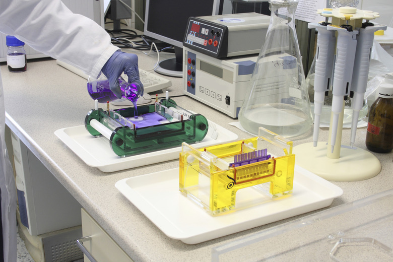
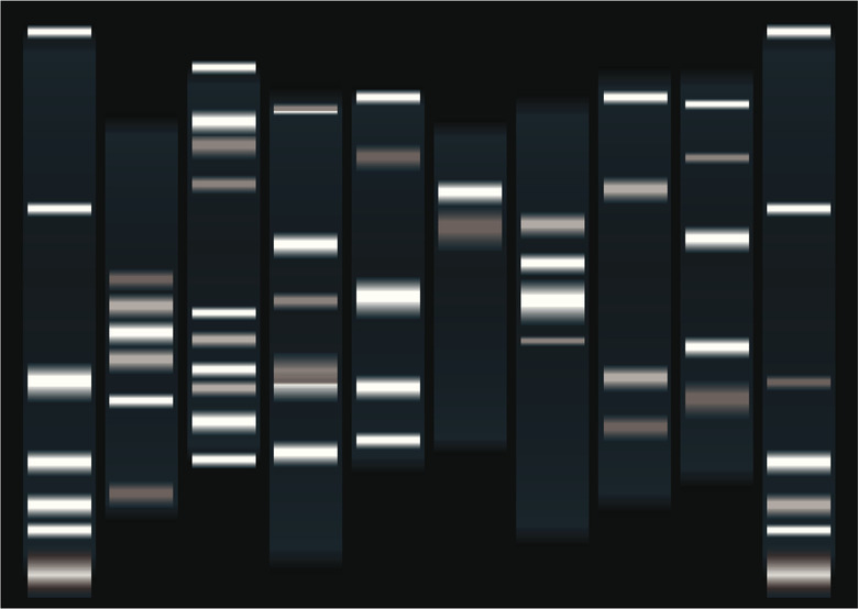
Gel electrophoresis involves the use of a gel usually made out of polymers such as agarose. The gel is immersed in a buffer solution that conducts an electric field. The DNA sample of interest is first fragmented using restriction enzymes and is then injected into the gel. When the electric field is turned on, the DNA fragments in the gel migrate toward the positive electrode. If the DNA fragments are of different sizes, then the migration times will be different for each size fragment. The fragments are then visualized using a dye or autoradiography and are visible as bands in the gel.
Contamination of the Sample
Contamination of the Sample
The major application of electrophoresis is as a tool for the analysis of DNA in molecular biology, but it is also used in forensics as a means of identifying samples from a crime scene. It is important that sources of errors in this technique be minimized in order to get accurate results. One source of error is contamination of the DNA sample. If there is foreign DNA in the sample, the gel will have more bands than would be found in a gel that contains only the purified sample.
Problems with the Gel, Current and Buffer
Problems with the Gel, Current and Buffer
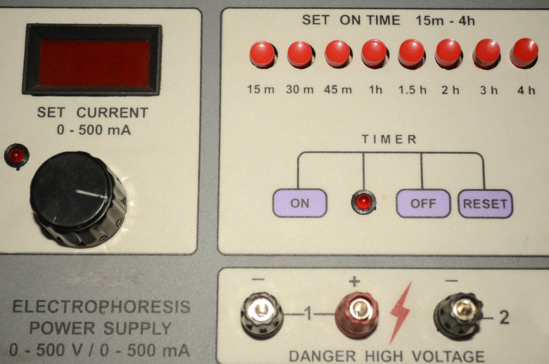
The concentration of the gel must also be correct to avoid errors. If the concentration is too high or too low, the fragments will migrate either too slowly or too quickly. This will lead to errors in resolving the different bands. During the electrophoresis run, care must be taken to ensure that the voltage is steady. Any fluctuations in the voltage will result in unsteady migration of DNA fragments, leading to errors in reading the bands. The buffer solution must also be of the correct composition, as a buffer with the wrong pH or ionic concentration will change the shape of the DNA fragments, also changing their migration times.
Proper Visualization
Proper Visualization
Most importantly, the gel must be visualized properly. If the concentration of the dye or radioactive probe used to visualize the samples is too high, the resulting image will be very messy, as residual fragments will also be visualized. If the gel concentration is too low, there will be no visualization. When the correct processes have been followed during all stages, gel electrophoresis will yield results that are accurate and can be used with great confidence. As with all scientific procedures, gel electrophoresis can be prone to errors, but these can be minimized with proper preparation and handling.
Cite This Article
MLA
Suico, Joshua. "Sources Of Error In Gel Electrophoresis" sciencing.com, https://www.sciencing.com/sources-of-error-in-gel-electrophoresis-12359700/. 28 September 2017.
APA
Suico, Joshua. (2017, September 28). Sources Of Error In Gel Electrophoresis. sciencing.com. Retrieved from https://www.sciencing.com/sources-of-error-in-gel-electrophoresis-12359700/
Chicago
Suico, Joshua. Sources Of Error In Gel Electrophoresis last modified August 30, 2022. https://www.sciencing.com/sources-of-error-in-gel-electrophoresis-12359700/
