The Structure & Function Of A Cell
Cells represent the smallest, or at least the most irreducible, objects that feature all of the qualities associated with the magical prospect called "life," such as metabolism (extracting energy from outside sources to power internal processes) and reproduction. In this respect, they occupy the same niche in biology as atoms do in chemistry: They can certainly be broken down into smaller pieces, but in isolation, those pieces cannot really do a whole lot. In any case, the human body certainly contains a lot of them – well over 30 trillion (that's 30 million million).
A common refrain in both the natural sciences and the engineering world is "form fits function." This essentially means that if something has a given job to do, it will probably look like it is capable of doing that job; conversely, if something appears to be made for accomplishing a given task or tasks, then there's a good chance this is exactly what that thing does.
The organization of cells and the processes they carry out are intimately related, even inseparable, and mastering the basics of cell structure and function is both rewarding in itself and necessary for fully understanding the nature of living things.
Discovery of the Cell
Discovery of the Cell
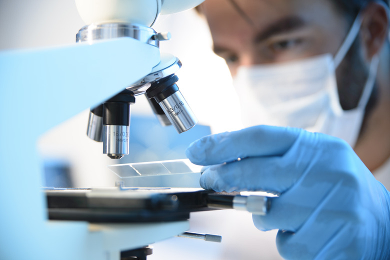
The concept of matter – both living and nonliving – as consisting of vast numbers of discrete, similar units has existed since the time of Democritus, a Greek scholar whose life spanned the 5th and 4th centuries B.C. But since cells are far too small to be seen with the unaided eye, it was not until the 17th century, after the invention of the first microscopes, that anyone was able to actually visualize them.
Robert Hooke is generally credited with coining the term "cell" in a biological context in 1665, although his work in this area focused on cork; about 20 years later, Anton van Leeuwenhoek discovered bacteria. It would be another several centuries, however, before the specific parts of a cell and their functions could be clarified and fully described. In 1855, the relatively obscure scientist Rudolph Virchow theorized, correctly, that living cells can only come from other living cells, even though the first observations of chromosome replication were still a couple of decades away.
Prokaryotic vs. Eukaryotic Cells
Prokaryotic vs. Eukaryotic Cells
Prokaryotes, which span the taxonomic domains Bacteria and Archaea, have existed for about three and a half billion years, which is about three-fourths the age of the Earth itself. (Taxonomy is the science dealing with the classification of living things; domain is the highest-level category within the hierarchy.) Prokaryotic organisms usually consist of only a single cell.
Eukaryotes, the third domain, include animals, plants and fungi – in short, anything alive that you can actually see without lab instruments. The cells of these organisms are believed to have arisen from prokaryotes as a result of endosymbiosis (from the Greek from "living together inside"). Close to 3 billion years ago, a cell engulfed an aerobic (oxygen-using) bacterium, which served the purposes of both life forms because the "swallowed" bacterium provided a means of energy production for the host cell while providing a supportive environment for the endosymbiont.
Read more about the similarities and differences of prokaryotic and eukaryotic cells.
Cell Composition and Function
Cell Composition and Function
Cells vary widely in size, shape and the distribution of their contents, especially within the realm of eukaryotes. These organisms are much larger as well as much more diverse than prokaryotes, and in the spirit of "form fits function" referenced previously, these differences are evident even at the level of individual cells.
Consult any cell diagram, and no matter what organism the cell belongs to, you are assured of seeing certain features. These include a plasma membrane, which encloses the cellular contents; the cytoplasm, which is a jelly-like medium forming most of the cell's interior; deoxyribonucleic acid (DNA), the genetic material that cells pass along to the daughter cells that form when a cell divides in two during reproduction; and ribosomes, which are structures that are the sites of protein synthesis.
Prokaryotes also have a cell wall external to the cell membrane, as do plants. In eukaryotes, the DNA is enclosed in a nucleus, which has its own plasma membrane very similar to the one surrounding the cell itself.
The Plasma Membrane
The Plasma Membrane
The plasma membrane of cells consists of a phospholipid bilayer, the organization of which follows from the electrochemical properties of its constituent parts. The phospholipid molecules in each of the two layers includes hydrophilic "heads," which are drawn to water because of their charge, and hydrophobic "tails," which are not charged and therefore tend to point away from water. The hydrophobic portions of each layer face each other on the interior of the double membrane. The hydrophilic side of the outer layer faces the exterior of the cell, while the hydrophilic side of the inner layer faces the cytoplasm.
Crucially, the plasma membrane is semipermeable, which means that, rather like a bouncer at a nightclub, it grants entry to certain molecules while denying entry to others. Small molecules such as glucose (the sugar that serves as the ultimate fuel source for all cells) and carbon dioxide can move freely in and out of the cell, dodging the phospholipid molecules aligned perpendicular to the membrane as a whole. Other substances are actively transported across the membrane by "pumps" powered by adenosine triphosphate (ATP), a nucleotide that serves as the energy "currency" of all cells.
Read more about the structure and function of the plasma membrane.
The Nucleus
The Nucleus
The nucleus functions as the brain of eukaryotic cells. The plasma membrane around the nucleus is called the nuclear envelope. Inside the nucleus are chromosomes, which are "chunks" of DNA; the number of chromosomes varies from species to species (humans have 23 distinct kinds, but 46 in all – one of each type from the mother and one from the father).
When a eukaryotic cell divides, the DNA inside the nucleus does so first, after all of the chromosomes are replicated. This process, called mitosis, is detailed later.
Ribosomes and Protein Synthesis
Ribosomes and Protein Synthesis
Ribosomes are found in the cytoplasm of both eukaryotic and prokaryotic cells. In eukaryotes they are clustered along certain organelles (membrane-bound structures that have specific functions, like organs such as the liver and kidneys do in the body on a larger scale). Ribosomes make proteins using instructions carried in the "code" of DNA and transmitted to the ribosomes by messenger ribonucleic acid (mRNA).
After mRNA is synthesized in the nucleus using DNA as a template, it leaves the nucleus and attaches itself to ribosomes, which assemble proteins from among 20 different amino acids. The process of making mRNA is called transcription, while protein synthesis itself is known as translation.
Mitochondria
Mitochondria
No discussion of eukaryotic cell composition and function could be complete or even relevant without a thorough treatment of mitochondria. These organelles that are remarkable in at least two ways: They have helped scientists learn a great deal about the evolutionary origins of cells in general, and they are almost solely responsible for the diversity of eukaryotic life by permitting the development of cellular respiration.
All cells use the six-carbon sugar glucose for fuel. In both prokaryotes and eukaryotes, glucose undergoes a series of chemical reactions collectively termed glycolysis, which generates a small amount of ATP for the cell's needs. In almost all prokaryotes, this is the end of the metabolic line. But in eukaryotes, which are capable of using oxygen, the products of glycolysis pass into the mitochondria and undergo further reactions.
The first of these is the Krebs cycle, which creates a small amount of ATP but mostly functions to stockpile intermediate molecules for the grand finale of cellular respiration, the electron transport chain. The Krebs cycle takes place in the matrix of the mitochondria (the organelle's version of a private cytoplasm), while the electron transport chain, which produces the overwhelming majority of ATP in eukaryotes, transpires on the inner mitochondrial membrane.
Other Membrane-Bound Organelles
Other Membrane-Bound Organelles
Eukaryotic cells boast a number of specialized elements that underscore the extensive, interrelated metabolic needs of these complex cells. These include:
- **Endoplasmic reticulum:** This organelle is a network of tubules consisting of a plasma membrane that is continuous with the nuclear envelope. Its job is to modify newly manufactured proteins to prepare them for their downstream cellular functions as enzymes, structural elements and so on, tailoring them for the cell's specific needs. It also manufactures carbohydrates, lipids (fats) and hormones. The endoplasmic reticulum appears as either smooth or rough on microscopy, forms that are abbreviated SER and RER respectively. The RER is so designated because it it "studded" with ribosomes; this is where the protein modification occurs. The SER, on other other hand, is where the aforementioned substances are assembled.
- **Golgi bodies:** Also called the Golgi apparatus. It looks like a flattened stack of membrane-bound sacs, and it packages lipids and proteins into vesicles that then break away from the endoplasmic reticulum. The vesicles deliver the lipids and proteins to other parts of the cell.
- **Lysosomes:** All metabolic processes generate waste, and the cell must possess a means of getting rid of it. This function is taken care of by lysosomes, which contain digestive enzymes that break down proteins, fats and other substances, including worn-out organelles themselves.
- **Vacuoles and vesicles:** These organelles are sacs that shuttle around various cellular components, taking them from one intracellular location to the next. The main differences is that vesicles can fuse with other membranous components of the cell, whereas vacuoles cannot. In plant cells, some vacuoles contain digestive enzymes that can break down large molecules, not unlike lysosomes do.
- **Cytoskeleton:** This material consists of microtubules, protein complexes that offer structural support by extending from the nucleus through the cytoplasm all the way out to the plasma membrane. In this respect, they are like the beams and girders of a building, acting to keep the whole dynamic cell from collapsing in on itself.
DNA and Cell Division
DNA and Cell Division
When bacterial cells divide, the process is simple: The cell copies all of its elements, including its DNA, while approximately doubling in size, and then splits in two in a process known as binary fission.
Eukaryotic cell division is more involved. First, the DNA in the nucleus is replicated while the nuclear envelope dissolves, and then the replicated chromosomes separate into daughter nuclei. This is known as mitosis, and consists of four distinct stages: prophase, metaphase, anaphase and telophase; many sources insert a fifth stage, called prometaphase, right after prophase. After that, the nucleus divides and new nuclear envelopes form around the two identical sets of chromosomes.
Finally, the cell as a whole divides in a process known as cytokinesis. When certain defects are present in the DNA thanks to inherited malformations (mutations) or the presence of damaging chemicals, cell division may proceed unchecked; this is the basis for cancers, a group of diseases for which there remains no cure, although treatments continue to improve to allow for vastly improved quality of life.
Cite This Article
MLA
Beck, Kevin. "The Structure & Function Of A Cell" sciencing.com, https://www.sciencing.com/structure-function-cell-5105947/. 30 July 2019.
APA
Beck, Kevin. (2019, July 30). The Structure & Function Of A Cell. sciencing.com. Retrieved from https://www.sciencing.com/structure-function-cell-5105947/
Chicago
Beck, Kevin. The Structure & Function Of A Cell last modified August 30, 2022. https://www.sciencing.com/structure-function-cell-5105947/
