What Are The Four Nitrogenous Bases Of DNA?
Deoxyribonucleic acid, or DNA, may be the most famous single molecule in all of biology. The discovery of its double-helix structure in 1953 catapulted James Watson and Francis Crick a Nobel Prize, and even among non-science nerds, DNA is widely known for playing a major part in the innumerable traits that are passed from parents to offspring. In the past few decades, DNA has also become noteworthy for its role in forensic science; "DNA evidence," a phrase that could not have meaningfully existed until at least the 1980s, has now become an almost obligatory utterance in crime and police-procedural television shows and motion pictures.
Beyond such mundane trivia, however, lies an elegant and impressively well-studied structure that exists in almost every cell of every living thing. DNA is the stuff of genes on a smaller scale and chromosomes, which are collections of many, many genes, on a larger scale; together, all of the chromosomes in an organism (humans have 23 pairs, including 22 pairs of "regular" chromosomes and a pair of sex chromosomes) are known as the organism's genome.
If you have ever taken a biology class or watched an educational program on basic genetics, even if you don't recall much of it, you probably remember something like this:
...ACCCGTACGCGGATTAG...
The letters A, C, G and T may be regarded the schematic cornerstones of molecular biology. They are abbreviations for the names of the four so-called nitrogenous bases found in all DNA, with A standing for adenine, C for cytosine, G for guanine and T for thymine. (For simplicity, these abbreviations will usually be employed throughout the remainder of this article.) It is specific combinations of these bases, in groups of three called triplet codons, that ultimately serve as the instructions for what proteins your body's cellular manufacturing plants make. These proteins, each of which is a product of a particular gene, determine everything from what foods you can and cannot digest easily, to the color of your eyes, your ultimate adult height, whether you can "roll" your tongue or not and many other traits.
Before a thorough treatment of each of these marvelous bases is given, a treatise on the basics of DNA itself is in order.
Nucleic Acids: Overview
Nucleic Acids: Overview
DNA is one of two nucleic acids found in nature, the other being RNA, or ribonucleic acid. Nucleic acids are polymers, or long chains, of nucleotides. Nucleotides include three elements: a pentose (five-atom-ring) sugar, a phosphate group and a nitrogenous base.
DNA and RNA differ in three basic ways. First, the sugar in DNA is deoxyribose, while that in RNA is ribose; the difference between these is that deoxyribose contains one fewer oxygen atom outside the central ring. In addition, DNA is almost always double-stranded, while RNA is single-stranded. Finally, while DNA contains the aforementioned four nitrogenous bases (A, C, G and T), RNA contains A, C, G and uracil (U) in place of T. This difference is essential in stopping the enzymes that act on RNA from exerting activity on DNA and conversely.
Putting this all together, a single DNA nucleotide therefore contains one deoxyribose group, one phosphate group and a nitrogenous base drawn from among A, C, G or T.
Some molecules that are similar to nucleotides, some of them serving as intermediates in the process of nucleotide synthesis, are important in biochemistry as well. A nucleoside, for example, is a nitrogenous base linked to a ribose sugar; in other words, it is a nucleotide missing its phosphate group. Alternatively, some nucleotides have more than one phosphate group. ATP, or adenosine triphosphate, is adenine linked to a ribose sugar and three phosphates; this molecule is essential in cellular energy processes.
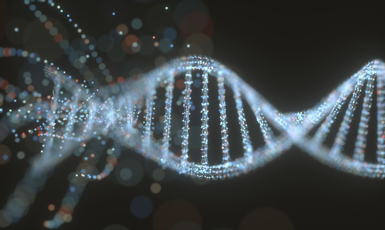
In a "standard" DNA nucleotide, deoxyribose and the phosphate group form the "backbone" of the double-stranded molecule, with phosphates and sugars repeating along the outer edges of the spiraling helix. The nitrogenous bases, meanwhile, occupy the interior portion of the molecule. Critically, these bases are linked to each other with hydrogen bonds, forming the "rungs" of a structure that, if not wound into a helix, would resemble a ladder; in this model, the sugars and phosphates form the sides. However, each DNA nitrogenous base can bind to one and only one of the other three. Specifically, A always pairs with T, and C always pairs with G.
As noted, deoxyribose is a five-atom-ring sugar. These four carbon atoms and one oxygen atom are arranged in a structure that, in a schematic representation, offers a pentagon-like appearance. In a nucleotide, the phosphate group is attached to the carbon designated number five by chemical naming convention (5'). the number-three carbon (3') is almost directly across from this, and this atom can bind to the phosphate group of another nucleotide. Meanwhile, the nitrogenous base of the nucleotide is attached to the 2' carbon in the deoxyribose ring.
As you may have gathered by this point, since the only difference from one nucleotide to the next is the nitrogenous base each includes, the only difference between any two DNA strands is the exact sequence of its linked nucleotides and hence its nitrogenous bases. In fact, clam DNA, donkey DNA, plant DNA and your own DNA consist of exactly the same chemicals; these differ only in how they are ordered, and it is this order that determines the protein product that any gene – that is, any section of DNA carrying the code for a single manufacturing job – will ultimately be responsible for synthesizing.
Exactly What Is a Nitrogenous Base?
Exactly What Is a Nitrogenous Base?
A, C, G and T (and U) are nitrogenous because of the large amount of the element nitrogen they contain relative to their overall mass, and they are bases because they are proton (hydrogen atom) acceptors and tend to carry a net positive electrical charge. These compounds do not need to be consumed in the human diet, although they are found in some foods; they can be synthesized from scratch from various metabolites.
A and G are classified as purines, while C and T are pyrimidines. Purines include a six-member ring fused to a five-member ring, and between them, these rings include four nitrogen atoms and five carbon atoms. Pyrimidines have only a six-member ring, which houses two nitrogen atoms and four carbon atoms. Each type of base also has other constituents projecting from the ring.
Looking at the math, it is clear that purines are significantly larger than pyrimidines. This explains in part why the purine A binds only to the pyrimidine T, and why the purine G binds only to the pyrimidine C. If the two sugar-phosphate backbones in double-stranded DNA are to remain the same distance apart, which they must if the helix is to be stable, then two purines bonded together would be excessively large, while two bonded pyrimidines would be excessively small.
In DNA, the purine-pyrimidine bonds are hydrogen bonds. In some instances, this is a hydrogen bonded to an oxygen, and in others it is a hydrogen bonded to a nitrogen. The C-G complex includes two H-N bonds and one H-O bond, and the A-T complex includes one H-N bond and one H-O bond.
Purine and Pyrimidine Metabolism
Purine and Pyrimidine Metabolism
Adenine (formally 6-amino purine) and guanine (2-amino-6-oxy purine) have been mentioned. Though not a part of DNA, other biochemically important purines include hypoxanthine (6-oxy purine) and xanthine (2,6-dioxy purine).
When purines are broken down in the body in humans, the end product is uric acid, which is excreted in the urine. A and G undergo slightly different catabolic (i.e., breakdown) processes, but these converge at xanthine. This base is then oxidized to generate uric acid. Normally, as this acid cannot be broken down further, it is excreted intact in urine. However, in some cases, an excess of uric acid can accumulate and cause physical problems. If the uric acid combines with available calcium ions, kidney stones or bladder stones can result, both of which are often very painful. An excess of uric acid can also cause a condition called gout, in which uric acid crystals are deposited in various tissues throughout the body. One way to control this is to limit intake of purine-containing foods, such as organ meats. Another is to administer the drug allopurinol, which shifts the purine breakdown pathway away from uric acid by interfering with key enzymes.
As for pyrimidines, cytosine (2-oxy-4-amino pyrimidine), thymine (2,4-dioxy-5-methyl pyrimidine) and uracil (2,4-dioxy pyrimidine) have already been introduced. Orotic acid (2,4-dioxy-6-carboxy pyrimidine) is another metabolically relevant pyrimidine.
The breakdown of pyrimidines is simpler than that of purines. First, the ring is broken. The end products are simple and common substances: amino acids, ammonia and carbon dioxide.
Purine and Pyrimidine Synthesis
Purine and Pyrimidine Synthesis
As stated above, purines and pyrimidines are made from components that can be found in abundance in the human body and do not need to be ingested intact.
Purines, which are synthesized mainly in the liver, are assembled from the amino acids glycine, aspartate and glutamate, which supply the nitrogen, and from folic acid and carbon dioxide, which provide the carbon. Importantly, the nitrogenous bases themselves never stand alone during the synthesis of nucleotides, because ribose enters into the mix before pure alanine or guanine appears. This produces either adenosine monophosphate (AMP) or guanosine monophosphate (GMP), both of which are nearly complete nucleotides ready to enter into a chain of DNA, although they can also be phosphorylated to produce adenosine di- and triphosphate (ADP and ATP) or guanosine di- and triphosphate (GDP and GTP).
Purine synthesis is an energy-intensive process, requiring at least four molecules of ATP per purine produced.
Pyrimidines are smaller molecules than purines, and their synthesis is correspondingly simpler. It occurs mainly in the spleen, thymus gland, gastrointestinal tract and testes in males. Glutamine and aspartate supply all of the required nitrogen and carbon. In both purines and pyrimidines, the sugar component of the eventual nucleotide is drawn from a molecule called 5-Phosphoribosyl-1-pyrophosphate (PRPP). Glutamine and aspartate combine to yield the molecule carbamoyl phosphate. This is then converted to orotic acid, which can then become either cytosine or thymine. Note that, in contrast to purine synthesis, pyrimidines destined for inclusion in DNA can stand as free bases (that is, the sugar component is added later). The transformation of orotic acid to cytosine or thymine is a sequential pathway, not a branched pathway, so cytosine is invariably formed first, and this can either be retained or further processed into thymine.
The body can make use of stand-alone purine bases apart from DNA synthetic pathways. Although purine bases are not formed during nucleotide synthesis, they can be incorporated midstream in the process by being "salvaged" from various tissues. This occurs when PRPP is combined with either adenosine or guanine from AMP or GMP plus two phosphate molecules.
Lesch-Nyhan syndrome is a condition in which the purine salvage pathway fails owing to an enzyme deficiency, leading to a very high concentration of free (unsalvaged) purine and therefore a dangerously high level of uric acid throughout the body. One of the symptoms of this unfortunate malady is that patients often display uncontrollable self-mutilating behavior.
Cite This Article
MLA
Beck, Kevin. "What Are The Four Nitrogenous Bases Of DNA?" sciencing.com, https://www.sciencing.com/what-four-nitrogenous-bases-dna-4596107/. 14 August 2018.
APA
Beck, Kevin. (2018, August 14). What Are The Four Nitrogenous Bases Of DNA?. sciencing.com. Retrieved from https://www.sciencing.com/what-four-nitrogenous-bases-dna-4596107/
Chicago
Beck, Kevin. What Are The Four Nitrogenous Bases Of DNA? last modified August 30, 2022. https://www.sciencing.com/what-four-nitrogenous-bases-dna-4596107/
