What Function Do Spindles Perform During Mitosis?
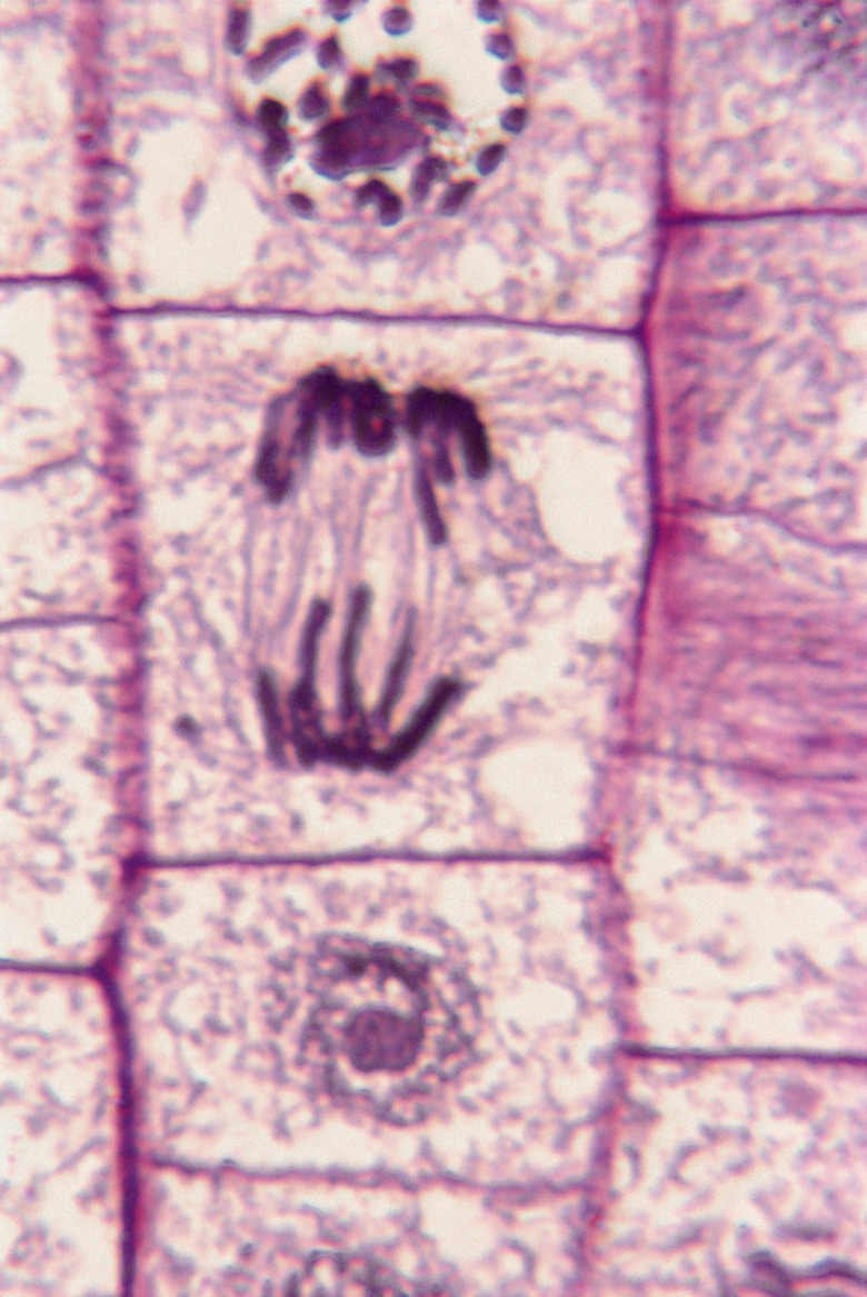
Spindle fibers are protein structures that form early in mitosis, or cell division. They consist of microtubules that originate from the centrioles, two wheel-shaped bodies located in the centromere area of the cell. The centromere is also known as the microtubule organizing center. The spindle fibers provide a framework and means of attachment that keep chromosomes organized, aligned and assorted during the entire process of mitosis, lessening the occurrence of aneuploidy, or daughter cells with incomplete sets of chromosomes. Aneuploidy is characteristic of cancers.
Components
Components
The spindle microtubules are protein fibers made up of as many as 45 different proteins that grow from the centrioles. They form a polymer, which is a large molecule made up of many similar molecules linked together. A number of proteins called molecular motors drive the spindle formation and functioning, including kinesins and dynein. Kinesins help establish the two opposite poles of the spindle, position the chromosomes between the poles and focus the spindle poles. Dynein regulates spindle length, spindle position and pole focusing and contributes to the checkpoint during metaphase. In metaphase, chromosome pairs line up along the midpoint of the dividing cell along the equatorial plane. Here they are checked for proper attachment to the spindle and readiness for separation during cell division.
Attachments
Attachments
Spindle microtubules attach to a specific protein complex called the kinetochore, which is in the centromere area near the center of each chromosome. Other microtubules attach to the chromosome arms or to the other end of the cell. The chromosomes can also create microtubules, as can the spindle itself. The spindle and chromosomal microtubule arrangement is a macromolecular machine that is intricate and dynamic.
Separation
Separation
Once the chromosomes have been checked at the equatorial plane, the adhesions between the two sets of chromosomes dissolve. This action allows the spindle fibers that attach the chromosomes to the centrioles at each end of the dividing cell to pull the two sets of chromosomes apart. Spindle microtubles that have grown into opposite sides of the cell originally have overlapping areas; but as the chromosomes begin to segregate during the anaphase stage of mitosis, the areas of overlap decrease and the cell elongates.
Segregation
Segregation
As anaphase proceeds, spindle fibers pull each set of chromosomes toward opposite ends of the dividing cell. Two methods of spindle shortening act to move the chromosomes. In one mechanism, the spindle fibers attached to the chromosomal kinetochores begin to quickly break down and depolymerize, which shortens the microtubules and moves the chromosomes closer to the pole to which the microtubles are attached. Another pulling mechanism occurs when motor proteins at the spindle poles pull the chromsomes closer. During the telophase stage of mitosis, each set of chromosomes segregates to the ends of the dividing cell, and the spindle fibers depolymerize and disappear, as do the centrioles. The cell then divides into two identical daughter cells.
References
- Scitable: Glossary: Spindle Fibers
- Uniprot: Subcellular Locations: Cellular Component Centrosome
- International Review of Cytology; Mechanisms of Mitotic Spindle Assembly and Function; C. E. Walczak and R. Heald
- Boundless: The Role of Mitotic Spindles in Cell Division
- TheFreeDictionary: Polymer
- Journal of Cellular Biology; Interzone Microtubule Behavior in Laate Anaphase and Telophase Spindles; W. M. Saxton and J. R. McIntosh
Cite This Article
MLA
Csanyi, Carolyn. "What Function Do Spindles Perform During Mitosis?" sciencing.com, https://www.sciencing.com/what-function-do-spindles-perform-during-mitosis-12731308/. 25 January 2014.
APA
Csanyi, Carolyn. (2014, January 25). What Function Do Spindles Perform During Mitosis?. sciencing.com. Retrieved from https://www.sciencing.com/what-function-do-spindles-perform-during-mitosis-12731308/
Chicago
Csanyi, Carolyn. What Function Do Spindles Perform During Mitosis? last modified March 24, 2022. https://www.sciencing.com/what-function-do-spindles-perform-during-mitosis-12731308/
