Prophase: What Happens In This Stage Of Mitosis & Meiosis?
The primary function of all living organisms, from the dispassionate stance of species survival, is to successfully propagate genetic material to subsequent generations. Part of this task, of course, is remaining alive and healthy for long enough to actually mate and reproduce. As a result of these realities, the fundamental units of living things, cells, have two primary jobs: making identical copies of themselves to maintain growth, perform repairs, and take care of other everyday functions at the level of tissues, organs and the whole organism; and generating specialized cells called gametes that combine with gametes from other organisms of the species to generate offspring.
The process of replicating whole cells to produce identical daughter cells as called mitosis, and it occurs in all eukaryotes, which are animals, plants and fungi (prokaryotes, almost all of which are bacteria, reproduce by binary fission, similar to mitosis but simpler). The generation of gametes occurs only in the gonads and is called meiosis. Both mitosis and meiosis are subdivided into five phases, which in the case of meiosis includes two rounds of each phase per original cell because meiosis results in four new cells rather than two. The first and longest of these phases is called prophase, which in meiosis I is further divided into five phases of its own.
What Is "Genetic Material"?
What Is "Genetic Material"?
All living things on Earth have DNA, or deoxyribonucleic acid, as their genetic material. DNA is one of a pair nucleic acids that exist in living systems, the other being ribonucleic acid (RNA). Both of these macromolecules – so named because they consist of large numbers of atoms, in this case arranged in long chains of repeating subunits called nucleotides – are absolutely critical, albeit in different ways. DNA, the root-level bearer of genetic information, is required to make RNA, but RNA comes in a variety of forms and is arguably more versatile.
The subunits from which both DNA and RNA are made are called nucleotides. Each of these consists of three parts: a five-carbon sugar that includes a central, pentagonal ring structure (in DNA this sugar is deoxyribose; in RNA it is ribose, which has one additional oxygen atom), a phosphate group and a nitrogenous (nitrogen-atom-rich) base. Each nucleotide has only one such base, but they come in four flavors for each nucleic acid. DNA features adenine (A), cytosine (C), guanine (G) and thymine (T); RNA includes the first three but substitutes uracil (U) for thymine. Because all of the variation between nucleotides is owed to differences in these bases, and nucleic acids consist of long chains of nucleotides, all of the variation between strands of DNA, and between DNA in different organisms, is owed to variation in these bases. Thus strands of DNA are written in terms of their base sequences, such as AAATCGATG.
DNA exists in living cells in the form of a double-stranded helix, or corkscrew shape. These strands are linked by hydrogen bonds between by their nitrogenous bases at each nucleotide; A uniquely pairs with T and C uniquely pairs with G, so if you know the sequence of one strand, you can easily predict the sequence of the other, called a complementary strand.
When messenger RNA (mRNA) is synthesized from DNA in a process called transcription, the mRNA made is complementary to the template DNA strand, and is thus identical to the strand of DNA not being used as a template except for U appearing in mRNA where T appears in DNA. This mRNA moves from the nucleus of cells where it is made to the cytoplasm, where it "finds" structures called ribosomes, which manufacture proteins using the mRNA's instructions. Each three-base sequence (e.g., AAU, CGC), called a triplet codon, corresponds to one of 20 amino acids, and amino acids are the subunits of whole proteins in the same manner that nucleotides are the subunits of nucleic acids.
Organization of DNA Within Cells
Organization of DNA Within Cells
DNA itself rarely appears in living things by itself. The reason for this, simply put, is the phenomenal amount of it that is required to carry the codes for all of the proteins an organism needs to make. A single, complete copy of your own DNA, for example, would be 6 feet long if stretched end to end, and you have a full copy of this DNA in almost every cell in your body. Since cells are only 1 or 2 microns (millionths of a meter) in diameter, the level of compression needed to pack your genetic material into a cell nucleus is astronomical.
The way your body does this is by studding your DNA with protein complexes called histone octamers to create a substance called chromatin, which is about two-thirds protein and one-third DNA. While adding mass to reduce size seems counterintuitive, think of it in roughly the same way as a department store paying security people to prevent loss of money through shoplifting. Without these comparatively heavy histones, which allow for highly extensive folding and spooling of DNA around their cores, DNA would have no means of being condensed. The histones are a necessary investment to this end.
Chromatin itself is divided into discrete molecules called chromosomes. Humans have 23 distinct chromosomes, with 22 of these being numbered and the remaining one being a sex chromosome (X or Y). All of your cells except gametes have two of every numbered chromosome and two sex chromosomes, but these are not identical, merely paired, because you receive one of each of these from your mother and the other from your father. Corresponding chromosomes inherited from each source are called homologous chromosomes; for example, your maternal and paternal copies of chromosome 16 are homologous.
Chromosomes, in newly formed cells, exist briefly in simple, linear form before replicating in preparation for cell division. This replication results in the creation of two identical chromosomes called sister chromatids, which are linked at a point called a centromere. In this state, then, all 46 of your chromosomes have been duplicated, making for 92 chromatids in all.
Overview of Mitosis
Overview of Mitosis
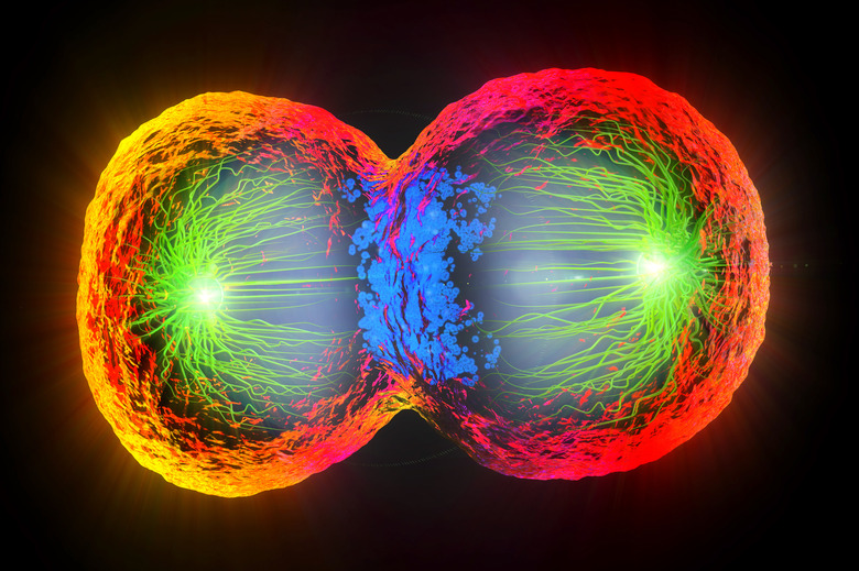
Mitosis, in which the contents of the nuclei of somatic cells (i.e., "everyday" cells, or non-gametes) divide, includes five phases: prophase, prometaphase, metaphase, anaphase and telophase. Prophase, discussed in detail shortly, is the longest of these and is chiefly a series of deconstructions and dissolutions. In prometaphase, all 46 chromosomes begin to migrate toward the middle of the cell, where they will form a line perpendicular to the direction in which the cell will soon be pulled apart. On each side of this line, called the metaphase plate, are structures called centrosomes; from these radiate protein fibers called microtubules, which form the mitotic spindle. These fibers connect to the centromeres of individual chromosomes on either side at a point called the kinetochore, engaging in a kind of tug of war to ensure that the chromosomes, or more specifically their centromeres, form a perfectly straight line along the metaphase plate. (Picture a platoon of soldiers going from standing in a recognizable rows and columns – a sort of "prometaphase" – to a rigid, inspection-ready formation – the equivalent of "metaphase.")
In anaphase, the shortest and most dramatic phase of mitosis, the spindle fibers pull the chromatids apart at their centromeres, with one chromatid drawn toward the centrosome on each side. The soon-to-divide cell now looks oblong under a microscope, being "fatter" on each side of the metaphase plate. Finally, in telophase, two daughter nuclei are completely formed by the appearance of nuclear membranes; this phase is like prophase run in reverse. After telophase, the cell itself divides in two (cytokinesis).
Overview of Meiosis
Overview of Meiosis
Meiosis unfolds in specialized cells of the gonads (testes in males, ovaries in females). In contrast to mitosis, which creates "everyday" cells for inclusion in existing tissues, meiosis creates gametes, which fuse with gametes of the opposite sex in fertilization.
Meiosis is divided into meiosis I and meiosis II. In meiosis I, instead of all 46 chromosomes forming a line along the metaphase plate as in mitosis, the homologous chromosomes "track down" each other and pair off, exchanging some DNA in the process. That is, maternal chromosome 1 links to paternal chromosome 1 and so on for the other 22 chromosomes. These pairs are called bivalents.
For each bivalent, the homologous chromosome from the father comes to rest on one side of the metaphase plate, and the homologous chromosome from the mother rests on the other. This occurs independently in each bivalent, so a random number of paternally sourced and maternally sourced chromosomes wind up on each side of the metaphase plate. The processes of DNA exchange (a.k.a. recombination) and random lining up (a.k.a. independent assortment) ensure diversity in the offspring because of the virtually unlimited range of DNA that results in gamete formation.
When the cell undergoing meiosis I divides, each daughter cell has one replicated copy of all 23 chromosomes, rather than 46 chromatids a la mitosis. All 46 centromeres are thus unperturbed at the onset of meiosis II.
Meiosis II is, for all practical purposes, a mitotic division, as the chromatids from meiosis I separate at the centromeres. The final outcome of both stages of meiosis is four daughter cells in two different identical pairs, each with 23 single chromosomes. This allows for the preservation of 46 chromosomes when male gametes (spermatocytes) and female gametes (ooctyes) join in fertilization.
Prophase in Mitosis
Prophase in Mitosis
Prophase occupies over half of mitosis. The nuclear membrane breaks down and forms small vesicles, and the nucleolus within the nucleus disintegrates. The centrosome divides in two, with the resultant components taking up residence on opposite sides of the cell. These centrosomes then begin generating microtubules that fan out toward the metaphase plate, similar, perhaps, to the way a spider generates its web. The individual chromosomes become fully compact, making them more recognizable under a microscope and allowing for easy visualization of the sister chromatids and the centromere between them.
Prophase in Meiosis
Prophase in Meiosis
Prophase of meiosis I includes five stages. In the leptotene phase, all of the structures of the not-yet-paired homologous chromosomes begin to condense, similar to what occurs in prophase in mitosis. In zygotene phase, the homologous chromosomes associate in a process called synapsis, with a structure called the synaptonemal complex forming between the homologs. In the pachytene phase, recombination between homologous chromosomes occurs (also called "crossing over"); think of this as you trading perhaps one sock and a hat with a sibling who you closely resemble in appearance and dress. In the diplotene phase, the bivalent begins to separate, but the homologs remain physically joined at their chiasmata. Lastly, in diakinesis, the chromosomes continue to pull farther apart, with the chiasmata moving toward their ends.
It is essential to recognize that without meiosis, and without the events of prophase I specifically, very little variation between different organisms would be evident. The shuffling of genetic material that occurs in this phase is the entire essence of sexual reproduction.
Prophase II, which occurs in the non-identical daughter cells formed by meiosis I, sees the individual chromosomes again condense into recognizable shapes, with the nuclear membrane dissolving as the mitotic spindle forms.
Cite This Article
MLA
Beck, Kevin. "Prophase: What Happens In This Stage Of Mitosis & Meiosis?" sciencing.com, https://www.sciencing.com/what-happens-in-prophase-13714454/. 24 September 2018.
APA
Beck, Kevin. (2018, September 24). Prophase: What Happens In This Stage Of Mitosis & Meiosis?. sciencing.com. Retrieved from https://www.sciencing.com/what-happens-in-prophase-13714454/
Chicago
Beck, Kevin. Prophase: What Happens In This Stage Of Mitosis & Meiosis? last modified August 30, 2022. https://www.sciencing.com/what-happens-in-prophase-13714454/
