Telophase: What Happens In This Stage Of Mitosis & Meiosis?
Cell division is an extremely important part in the development of all the cells of all organisms, including humans, animals and plants. Telophase is the last stage of cell division before cytokinesis occurs to split the cells into daughter cells. Mitosis is the cell division of all tissues and organs in which two identical daughter cells are produced. Sex cells divide in meiosis to produce four daughter cells that each contains only half of the number of chromosomes of the parent cell.
What Are Some Telophase Characteristics in Cell Division?
What Are Some Telophase
Characteristics in Cell Division?
In mitosis, or the division of cells in organisms other than sex cells, which are also called autosomes, telophase facts include the chromosomes moving to opposite ends of the new cell to form two identical nuclei. After the cell splits into two daughter cells, they will both be identical to the original parent cell in every manner.
In meiosis, or the division of cells in sex cells, the original parent cell duplicates and then divides twice, much in the same manner as in mitosis. However, the end product is four daughter cells that each contains only half the number of chromosomes. The reason they have only half the number of chromosomes is because the diploid cell, or parent cell, duplicates once and then divides twice to produce daughter cells that are haploid. Haploid actually means "half."
What Are the Stages of a Cell in Mitosis?
What Are the Stages of a Cell
in Mitosis?
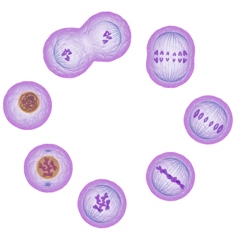
The acronym for the stages of a cell in the division process of mitosis is PMATI. It stands for prophase, metaphase, anaphase, telophase and interphase. In each stage of division the cell goes through distinct changes to create two identical daughter cells that can grow and heal wounds in the human body and animals.
Prophase is the next stage in the process when a cell receives signals telling it to divide. It is characterized by the cell duplicating the DNA and preparing itself for the actual cell division.
Metaphase in the stage in which all the pieces of the new cells have aligned their DNA along a central axis within the cell. The pair of centrioles, or organelles that specialize in cell division, move to the opposite ends or poles of the cell. The centrioles have fibers that connect to the DNA, and the DNA chromatin condenses to form chromosomes.
Anaphase is when the separation begins, and the chromosomes are pulled to opposite ends of the cell, readying for the division.
Telophase mitosis is the next stage in which the cell membrane splits the cell into two duplicate daughter cells.
Interphase is when a parent cell is in a resting state, and the phase that a cell remains in for the most part until dividing. The cell gains energy, grows and then duplicates the nucleic acids in preparation for the next cell division.
What Are the Stages of a Cell in Meiosis?
What Are the Stages of a Cell
in Meiosis?
The cell division process in meiosis is found in all organisms that can reproduce sexually including humans, plants and animals. Meiosis is a two-part division of cells to produce four daughter cells with half the number of chromosomes as the original or parent cell. The two-part division process is called meiosis I and meiosis II. So, telophase meiosis is characterized by telophase I and telophase II, just as all of the other stages occur twice in the process of division in meiosis.
The interphase stage is when a cell is in a resting state and gaining the items it will need for an upcoming cell division. This is the stage where cells remain for most of their life. The interphase is broken down into three phases, G1, S and G2. In the G1 phase, the cell increases in mass to prepare for division. The G represents gap and the one is the first phase, meaning that the G1 phase is the first gap phase in cell division of meiosis.
The S phase is the next stage when DNA is synthesized. S stands for synthesis. The G2 phase is the second gap phase in which the cell synthesizes its proteins, and it continues to increase in size. At the end of the interphase, the cell has nucleoli present, and the nucleus is bound by the nuclear envelope. The cell's chromosomes divide and are in the form of chromatin. In animal and human cells, the two pairs of centrioles form and are located on the outside of the nucleus.
Prophase I is the stage when several changes in the cell take effect. The chromosomes condense in size, and they then attach to the nuclear envelope. A pair of identical or homologous chromosomes line up closely to each other to form a tetrad, which is composed of four chromatids. This is known as synapsis. Crossing over may occur to create new genetic combinations that are different from either of the parent cells.
The chromosomes thicken, and then they detach from the nuclear envelope. The centrioles move away from each other and start migrating to opposite sides or poles of the cell. The nucleoli and the nuclear envelope break down, and the chromosomes start moving to the metaphase plate.
Metaphase I is the next stage in which the tetrads align at the metaphase plate in the cell, and the identical chromosome pairs or centromeres are now at the opposite sides of the cell.
In anaphase I, fibers develop from the opposite poles of the cell to pull the chromosomes toward the two poles. The two identical copies of a chromosome that are connected by a centromere, or sister chromatids, remain together after the chromosomes move to the opposite poles.
The next stage is telophase I, in which the spindle fibers continue pulling the homologous chromosomes to the opposite poles. After they reach the poles, each of the two poles contain a haploid cell, which contains half as many chromosomes as the parent cell. The division of cytoplasm usually occurs in telophase I. At the end of telophase I and the process of cytokinesis when the cell divides, each cell will have half the chromosomes of the parent cell. The genetic material does not duplicate again, and the cell moves into meiosis II.
In prophase II, the nuclei and the nuclear membrane break up as the spindle network of fibers appear. The chromosomes again begin to migrate to the metaphase II plate, which is at the center or the cell equator.
Metaphase II is the stage in which a cell's chromosomes align themselves at the metaphase II plate at the center of the cell and the fibers of the sister chromatids are pointing to the two opposite poles on opposite sides of the cell.
Anaphase II is the next stage of cell division in meiosis in which the sister chromatids separate from each other and start moving to the opposite ends of the cell. The spindle fibers that are not connected to the two chromatids lengthen, and this elongates the cell. The separation of the sister chromatids in a pair is the point when the chromatids become chromosomes, called daughter chromosomes. The cell poles move further apart as the cell elongates. At the end of this stage, each pole contains a complete set of chromosomes.
In telophase II, two distinct nuclei begin to form at the opposite poles of the cell. The cytoplasm divides through cytokinesis to form two distinct cells, which are called daughter cells, each with one-half the number of chromosomes as the parent cell. The end product after both stage I and II of meiosis is four daughter cells that are haploid. When haploid cells unite during fertilization of a sperm cell and an egg, they will become a diploid cell, just as the original parent cell was in the beginning of the cell before division.
What Is Chromosomal Non-Disjunction in Meiosis?
What Is Chromosomal
Non-Disjunction in Meiosis?
In normal cell division through meiosis, the division creates gametes or sex cells of eggs and sperm. There can be errors in the process that lead to mutations in gametes. Defective gametes can lead to a miscarriage in humans, or it can lead to genetic disorders or diseases, just as in the cell division of mitosis. Chromosomal non-disjunction is the result of the wrong number of chromosomes in a cell.
A normal gamete contains a total of 46 chromosomes because they get 23 chromosomes from each of the two parent's DNA. In meiosis I, the cell divides to produce two daughter cells, and in meiosis II, it divides again to produce four daughter cells that are haploid, containing half of the number of chromosomes of the original cell before division occurs. Human egg and sperm cells each have 23 chromosomes, so when fertilization occurs between sperm and an egg, it produces a cell with 46 chromosomes to produce a healthy baby.
Non-disjunction may occur when the chromosomes do not separate properly when the cell divides, so it creates gametes with the wrong number of chromosomes. A sperm or egg cell may have an extra chromosome, totaling 24, or it may be missing a chromosome, totaling 22. In human sex cells, this abnormality would become a baby with 45 or 47 chromosomes instead of the normal amount of 46. Non-disjunction can lead to a miscarriage, stillbirth or a genetic disorder.
The non-disjunction of autosomes, or the non-sex chromosomes, results in a miscarriage or a genetic disorder. Autosome chromosomes are numbered 1 through 22. In this case, the baby will have one extra chromosome or trisomy, meaning three chromosomes. Three copies of chromosome 21 produces a child with Down syndrome. Trisomy 13 causes Patau Syndrome, and trisomy 18 produces Edward's Syndrome. Other chromosomes that get an extra one will lead to babies that are rarely carried to term as in the chromosomes 15, 16 and 22.
The non-disjunction of the sex cells on chromosome number 23 produce less drastic results that in the autosomes. Normally, males have the sex chromosome combination of XY, and females have the combination of XX in a normal cell. If a male or female gains an extra sex chromosome or loses a sex chromosome, it can lead to genetic disorders, with some being more serious than others or with no effects on the baby.
Klinefelter Syndrome occurs when a male has an extra X chromosome or the combination of XXY. A male that gains an extra Y chromosome, expressed as the chromosome combination of XYY also causes Klinefelter Syndrome. A female that is missing one X chromosome or only has one copy of X causes Turner's syndrome. This combination in females is the only case in a missing sex chromosome that produces a female baby that can survive without the other X chromosome. If a female receives an extra X or has trisomy X, expressed as a chromosome combination of XXX, the female baby will have no symptoms of any sort.
Cite This Article
MLA
Lougee, Mary. "Telophase: What Happens In This Stage Of Mitosis & Meiosis?" sciencing.com, https://www.sciencing.com/what-happens-in-telophase-13714452/. 5 September 2018.
APA
Lougee, Mary. (2018, September 5). Telophase: What Happens In This Stage Of Mitosis & Meiosis?. sciencing.com. Retrieved from https://www.sciencing.com/what-happens-in-telophase-13714452/
Chicago
Lougee, Mary. Telophase: What Happens In This Stage Of Mitosis & Meiosis? last modified March 24, 2022. https://www.sciencing.com/what-happens-in-telophase-13714452/