What Is A Gamete?
Gametes, also called sex cells or germ cells, are unique among the many types of cells in your body for having only 23 chromosomes, half the number that your other cells have. Everyday cells in tissues throughout your body have two copies of every chromosome, one from each of your parents. Human chromosomes are numbered 1 through 22, with the remaining chromosome, a sex chromosome, assigned a letter instead of a number – "X" or "Y." Matched copies of chromosomes – that is, chromosomes with the same assigned number, like chromosome 11 or chromosome 18 – are called homologous chromosomes, and they look the same under a microscope even if they differ at the level of their precise DNA composition. That is, the copy of chromosome 9 you received from your mother looks like the copy of chromosome 9 you received from your father, and so on for the other chromosomes.
As you may have guessed or learned from previous research, your everyday cells have one whole copy of the DNA supplied by the chromosomes of each of your parents because, about nine months before you were born, a cell from your mother and a cell your father joined together to create the cell that ultimately became the person you are now. But if each of those cells from your parents had carried 46 chromosomes, like most human cells do, your cells would have 92. The unique process of gamete formation in meiosis is what both preserves chromosome number across generations and ensures genetic diversity, a trait that is vital for the survival of any species.
Cell Division Basics
Cell Division Basics
Deoxyribonucleic acid (DNA) serves as the genetic material material in all living things. ("Genetic material" in this context refers to a complete set of chemically coded information that can be passed to offspring, i.e., is heritable.) In prokaryotes, a group for all intents and purposes synonymous with bacteria, this genetic information usually exists in the form of a ring, meaning that bacteria possess a single circular chromosome (more on these structures soon). This DNA is not part of a nucleus, for prokaryotes do not have internal organelles enclosed by double plasma membranes.
Eukaryotic organisms (plants, animals and fungi) have DNA enclosed in a double membrane, forming the nucleus that is unique to eukaryotic cells. The DNA of eukaryotes is divided into discrete chunks called chromosomes, which are also packaged with distinct structural proteins. As touched on above, humans cells, gametes excepted, have 46 chromosomes. Eukaryotic organisms also possess mitochondria, cigar-shaped organelles that are believed to have functioned over a billion years ago as free-standing bacteria in their own right; these are involved in aerobic respiration, but also possess their own DNA.
DNA, in addition to being the feature presentation of chromosomes, is is functionally divided into genes, which are lengths of DNA that carry the code for one specific protein product. In a process called transcription, DNA is used as a template to synthesize a similar molecule called messenger RNA (mRNA). This molecule then migrates out of the nucleus (in eukaryotes) and to the ribosomes that sit in the cell cytoplasm. Here, mRNA is used to manufacture proteins from amino acids in a process called translation.
More to the point if this discussion, DNA also undergoes replication, which simply means that it makes a copy of itself. Each cell's DNA does this in its entirety exactly once as a precursor to cell division. That is, in humans, all 46 human chromosomes, each of which contains a single very long DNA molecule, replicate before cell division can occur.
Bacterial cell division is often called binary fission and involves the single-celled organism simply dividing in two to make a pair of copies identical to the parent organism. Binary fission is a form of asexual reproduction, meaning that no mixing of genetic material between different bacteria occurs as part of the normal reproductive process. Eukaryotic cell division, on the other hand, takes two forms. In mitosis, the process is very much like that of bacterial fission, albeit more complicated owing to the greater complexity of eukaryotic cells. In meiosis, however, the mechanism is subtly yet powerfully different.
Gamete Cells
Gamete Cells
Gametes are produced in the gonads of animals – testes in men and ovaries in women. Also called sex cells or germ cells, these gametes go by different names in different organisms. In males, the gametes are called spermatocytes, while in females they are known as oocytes.
Gametes, as noted, have one copy of every numbered chromosome and one sex chromosome. Each of these chromosomes is a mosaic, or patchwork, of the material in the corresponding chromosomes of the organism's mother and father. That is, the copy of chromosome 14 that sits in any of the gametes your own body produces represents a blend of the material from the copy of chromosome 14 you inherited from your father and the material from the copy of chromosome 14 you inherited from your mother, and similarly for the rest of your chromosomes. Moreover, each gamete your gonads produce is a unique blend of your maternal and paternal chromosomes. If this were not the case, all of the children resulting from the union of a given couple would look exactly the same because each child would result from the fusion of genetically indistinguishable gametes. This implies that the formation of individual gametes, called gametogenesis, includes one or more steps that operate with some degree of randomness. In fact, there are two such distinct steps, explored in a subsequent section.
Chromosomes
Chromosomes
Before undertaking a description of gamete formation, it is useful to explore chromosomes in more detail as these are what ultimately get taken apart, shuttled around and reassembled during cell reproduction.
Chromosomes consist of distinct segments of chromatin, which in eukaryotes is material consisting of a mixture of DNA and proteins called histones. Histones cluster together in groups of eight subunits called octamers, and the DNA in the associated chromatin winds itself around each histone octamer like thread wraps its way around a spool, making about two revolutions per octamer. This condenses the chromatin from its linear form to some degree, but it is the successive stacking of these DNA-octamer complexes, called nucleosomes, that really allows chromatin to be super-condensed. An entire copy of your DNA sites in every one of your cells, yet stretched out in a straight line, this DNA would reach to 6 feet in length.
Your 23 pairs of chromosomes do not contain equal amounts of chromatin, and they vary considerably in size. When DNA replicates, each chromosome remains bound at a fixed position to the just-made copy. This point is called a centromere, and the two identical copies of each chromosome are called sister chromatids. The centromere, despite its name, is not usually in the middle of the chromatids it links, but toward one end – making it easier to distinguish individual numbered chromosomes from each other under a microscope. The shorter chromatid portions on one end of the centromere are called the p-arms, while the longer arms are called the q-arms.
Gametogenesis: Mitosis Versus Meiosis I and II
Gametogenesis: Mitosis Versus Meiosis I and II
Mitosis is the term for cell division that produces daughter-cell DNA identical to the parent and to each other. Meiosis, on the other hand, results in daughter cells that are genetically unique and distinct from each other.
Shortly before mitosis, which is divided for convenience into four phases (prophase, metaphase, anaphase and telophase), the cell chromosomes, which usually sit in a loose cluster like carelessly tossed-aside yarn, replicate (until this point, each one exists as a single linear chromatid) and begin to condense into their characteristic shapes. They then migrate toward the middle of the cell and assemble themselves in a line of 46, with the ends of one set of chromatids adjoining the ends of those on the next chromosome. Microtubules extending perpendicularly from the line formed by the chromosomes attach themselves to the sides of the chromosomes and pull them apart, so that each newly forming daughter cell receives one sister chromatid from each of the 46 chromosomes. The cell finishes dividing and forms new membranes around the new nuclei and the two new cells as a whole.
In meiosis, the process begins with a complete replication of DNA of all 46 chromosomes, as in mitosis. In the testicular and ovarian cells targeted for gamete production, however, the way chromosomes line up along the axis of division is much different. In meiosis I, the homologous chromosomes "find" each other and bind to produce a structure with two side-by-side chromosomes, one from the mother and one from the father, called a bivalent. With the homologous chromosomes touching, they trade portions of their DNA with each other. For example, a given amount of DNA on the long arm of the mother's copy of chromosome 6 (labeled q6) might find its way into the corresponding spot on the father's chromosome, and accept the father's section of q6 in its place. This is called crossing over, and is one of two main factors propelling the genetic diversity that results from meiosis.
Also, when the bivalents line up along the line of cell division, the mother's duplicated chromosome is on one side, while the father's is on the other. However, which one lands on which side is completely random with respect to all of the other 22 chromosomes. This is referred to as independent assortment and also contributes mightily to genetic diversity in sexually reproducing organisms. In fact, the number of possible bivalent arrangements is 2 raised to the 23rd power – some 8.4 million different combinations.
When this cell splits, completing meiosis I, the result is two non-identical cells containing 23 pairs of chromatids joined at their centromeres. These chromatids, however, while very similar, are not sister chromatids, owing to the phenomenon of crossing over in meiosis I detailed above. These two daughter cells then immediately undergo another cell division, this one resembling mitosis in that chromatids are pulled apart at the centromeres and separated. Recall, though, that this line of dividing chromosomes is only 23 in number, not 46, because of the way the chromosomes pair off in meiosis I. This means that each of the four daughter cells resulting from meiosis has 23 chromosomes, the human haploid number. 46 is considered the diploid number.
A Brief Note on Oogenesis and Spermatogenesis
A Brief Note on Oogenesis and Spermatogenesis
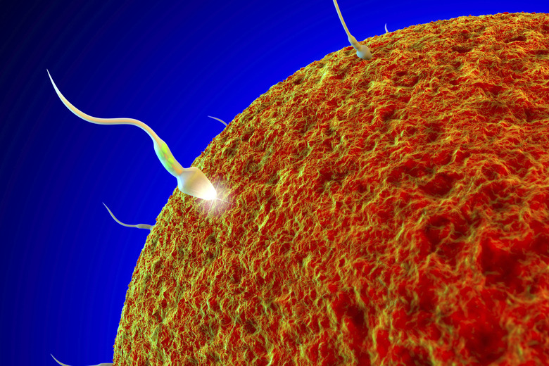
Spermatozoa, the flagella-bearing and "swimming" sperm that carry spermatocytes, are clearly different from egg cells. Correspondingly, gamete formation in males (spermatogenesis) is different from that in females (oogenesis). For example, each meiosis in females results in one daughter cell, not four as in spermatogenesis. Meiosis in females is initiated just once in the course of a woman's lifetime, with the resulting ooctyes reaching maturation about once every 28 days over the course of a woman's fertile lifetime. Spermatocytes, in contrast, repeatedly undergo the mitosis-like divisions of meiosis II to produce a far greater abundance of total gametes over a male's lifetime.
Cite This Article
MLA
Beck, Kevin. "What Is A Gamete?" sciencing.com, https://www.sciencing.com/what-is-a-gamete-13714445/. 17 September 2018.
APA
Beck, Kevin. (2018, September 17). What Is A Gamete?. sciencing.com. Retrieved from https://www.sciencing.com/what-is-a-gamete-13714445/
Chicago
Beck, Kevin. What Is A Gamete? last modified August 30, 2022. https://www.sciencing.com/what-is-a-gamete-13714445/