What Is A Zygote?
Sexual reproduction, which both plants and animals carry out, involves the fusion of gametes, or sex cells, to form a zygote, the technical term for what most people refer to as "a fertilized egg" in everyday language. Sexual reproduction seems like a cumbersome thing, biologically and energetically speaking, compared to what bacteria do – simply divide in two to make a pair of perfect new copies of the parent organism. But without this form of reproduction, a species could not experience genetic variation through random mixing of parental DNA; all offspring would be identical and thus identically vulnerable to environmental threats such as predators, extreme weather and microbial diseases. This would negatively affect species survival and hence is not an evolutionarily helpful way to reproduce in the long term, even if it is simple and reliable.
Zygotes pass through a series of phases en route to becoming full-fledged versions of their parents. Before undertaking a basic study of embryology, however, it is useful to know how sexual reproduction at the cellular level works and how it ensures genetic diversity. This requires basic knowledge of nucleic acids, chromosomes and genes, and cell division before the formation of zygotes can be adequately explored.
Nucleic Acids: the Basis of Life
Nucleic Acids: the Basis of Life
Deoxyribonucleic acid (DNA) has achieved great notoriety since its double-helix structure was famously elucidated in 1953 by a team of researchers including James Watson, Francis Crick and Rosalind Franklin. Anyone who watches police procedural shows or films these days knows that human DNA can be used to uniquely identify people, like microscopic versions of fingerprints; most high-school graduates are likely aware that DNA, in a tangible sense, makes us who we are and also reveals a great deal about both our parents and any children we have, presently or in the future.
DNA, in fact, is the stuff of which genes are made. A gene is simply a length of DNA molecule that carries the biochemical code for making a particular protein product, such as an enzyme or collagen fiber. DNA is a macromolecule that consists of monomers called nucleotides, each of which in turn has three components: a five-carbon sugar (deoxyribose in DNA, ribose in RNA), a phosphate group and a nitrogen-rich base. Variation in nucleotides results from variation in these nitrogenous bases, as DNA and RNA each have four kinds – adenine (A), cytosine (C), guanine (G) and thymine (T). (In RNA, uracil, or U, is substituted for T.) Consequently, unique strands of DNA are produced by the novel sequences of DNA they contain. For example, a strand with the nucleotide sequence ATTTCGATTA might hold the code for one gene product, while TAGCCCGTATT might hold the code for another. (Note: These are randomly selected sequences.
Because DNA is double-stranded, each base pairs up with a base on the complementary strand in a strict way: A always with T, and C always with G. Thus the strand ATTTCGATTA would pair with the strand TAAAGCTAAT under these inviolable rules.
DNA is believed to be the largest single molecule in the body, ranging up to many millions of base pairs (sometimes expressed as nucleotides) in length. Each individual chromosome, in fact, consists of one very long DNA molecule along with a significant amount of structural protein.
Chromosomes
Chromosomes
Every living cell in your body includes a nucleus, as do those of every other eukaryote (e.g., plants, animals and fungi), and within that nucleus is DNA bundled with proteins to create a material called chromatin. This chromatin, in turn, is chopped up into discrete units called chromosomes. Humans have 23 distinct chromosomes, including 22 numbered chromosomes (called autosomes) and one sex chromosome. Females have two X-chromosomes, while males have one X-chromosome and one Y-chromosome. In a sense, then, the father in any mating union "determines" the sex of the offspring.
Chromosomes are found in pairs in all cells with the exception of gametes, to be discussed in detail shortly. This means that when a typical cell divides, it creates two identical daughter cells, each with only one copy of each chromosome. Each of these 23 chromosomes soon replicates (i.e., makes a copy it itself), returning the number of chromosomes in ordinary cells to 46 again. This division of cells to create two identical cells is called mitosis, and it is both how your body replenishes dead and worn-out calls throughout the body and how single-celled organisms such as bacteria reproduce and "birth" entire copies of themselves.
Chromosomes, in the replicated state, consist of two identical halves called chromatids, which are joined by a condensed spot of chromatin called the centromere. So, while a single chromosome is a linear entity, a replicated chromosome looks more like an asymmetrical letter "X," or a pair of boomerangs meeting at the apices of their curve. Despite its name, the centromere is not usually centrally located, making for lopsided chromosomes. The material on the side of the centriole that appears smaller represents the p-arms of the two identical chromatids, while the other side side includes the q-arms.
The reproduction of gametes resembles mitosis in many respects, but the bookkeeping of genetic material can be confusing, and the seemingly superficial differences between mitosis and meiosis are why you, and only you, look exactly like you among the billions of people alive today (unless you have an identical twin, that is).
Meiosis I and II
Meiosis I and II
Gametes, or sex cells – sperm cells in human males and ova (eggs) in females – have only one copy of each chromosome, or 23 chromosomes in all. Gametes are produced in germ cells, where meiosis takes place in two stages, meiosis I and meiosis II.
At the start of meiosis I, the germ cell contains 46 chromosomes in 23 pairs, just like regular (somatic) cells do at the start or mitosis. However, in meiosis, the chromosomes are not pulled apart in a way such that each daughter cell receives one chromatid from every chromosome, for example, one from the maternally contributed copy of chromosome 1, one from the paternally contributed copy of chromosome 1, and so on. Instead, the homologous chromosomes (i.e., chromosome 8 from the mother and chromosome 8 from the father) come into physical contact with each other, with their corresponding arms exchanging random amounts of material. Then, before the cell actually divides, the chromosomes randomly align themselves along the plane of division so that some daughter cells receive, say, 10 chromatids from the mother and 13 from the father, while the other daughter cell gets 13 and 10. These two processes unique to meiosis are called recombination and independent assortment, and if you like, think of them together as the thorough shuffling of a deck of 23 pairs of cards. The point, again, is to ensure genetic diversity thanks to a never-before-seen genome in every gamete.
Meiosis II begins with 23 chromosomes (or single chromatids, if you prefer) in each of two non-identical daughter cells. Meiosis II is unremarkable compared to meiosis I, and resembles mitosis in that it produces two identical daughter cells. At the end of the cell division in meiosis II, the original cell with 46 chromosomes has given rise to four cells in two identical pairs with 23 chromosomes each. These are gametes, the cells that go on to form zygotes.
Formation of the Zygote
Formation of the Zygote
In humans, zygotes are formed when a male gamete, formally called a spermatozoon, fuses with a female gamete, called an oocyte. This process is called fertilization. While you have perhaps heard of something called "the moment of conception," this is a colloquialism with no scientific content, for fertilization (conception) is no instantaneous process, though it is poignant to watch under a microscope or on film.
In humans, the head of the sperm cells undergo a process called capacitation that changes the glycoproteins in their coats, and in a sense prepares them for battle by making them more prepared to penetrate the outside of the oocyte. Like most of the early travelers who attempted to reach the South Pole or summit Mount Everest, only a small fraction of sperm introduced into the female reproductive tract even make it to the vicinity of the egg inside the female's uterus.
The sperm that winds up being the "lucky" carrier of the material that ultimately becomes part of the zygote forces its way through the outer wall of the oocyte, called the corona radiata, by both physical means (propulsion by the sperm's propeller-like flagella appendage, tantamount to swimming) and chemical means (the sperm secretes an enzyme called hyaluronidase that helps break down proteins in the corona radiata).
At this point, the sperm has actually done only part of the work required to serve as a zygote component. Inside the zona radiata of the egg cell is another coat, called the zona pellucida. Now the head of the sperm undergoes what is knows as an acrosome reaction, dumping a number of corrosive chemicals so as to dissolve this new layer and allow the sperm to drill its way into the oocyte interior. Exhausted, the sperm releases its chromosomes into the interior of the egg cell, while its outer membrane fuses with that of the egg cell. The head, tail and remaining contents of the sperm all fall away and disintegrate. This is why all of the mitochondria in the zygote come from the mother, a finding that has implications in the tracing of humans back to their remote ancestors.
When the gametes have physically come together, they each have nuclei of their own, each with 23 single-strand chromosomes. The sperm can contain either an X-chromosome or a Y-chromosome, but the egg will always contain an X-chromosome. When the sperm and egg themselves fuse together, this starts with the cytoplasm and the sharing of a single cell membrane, leaving two separate nuclei in the center. These nuclei, at this very early stage of the zygote, are called pronuclei. Once these have fused to form a single nucleus, the nascent organism is now officially a zygote.
Zygote vs. Embryo
Zygote vs. Embryo
The various stages of embryonic development are often used interchangeably. At times, this is justified; in truth, there is no firm division between, for example, an embryo and a fetus. Nevertheless, conventional terminology is helpful.
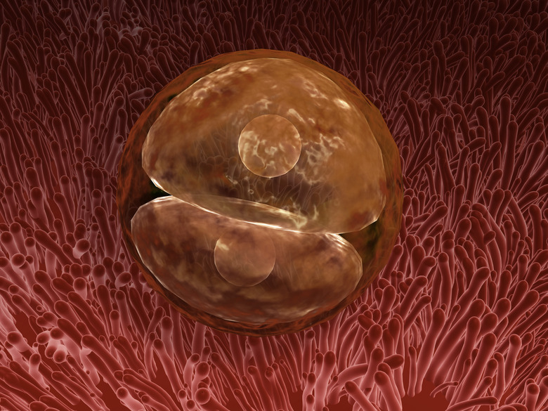
After the zygote is formed, the now-diploid (that is, containing 46 chromosomes) cell begins to divide. These early divisions are mitotic divisions, producing identical cells, and each takes about 24 hours. The cells thus formed are called blastomeres, and they actually become successively smaller with each division, preserving the overall size of the conceptus. At the end of six divisions, which leaves 32 total cells, the entity can be regarded as an embryo, specifically a morula (Latin for "mulberry"), a solid ball consisting of an inner cell mass, which eventually becomes the fetus itself, and an outer cell mass, which develops into the placenta.
Cite This Article
MLA
Beck, Kevin. "What Is A Zygote?" sciencing.com, https://www.sciencing.com/what-is-a-zygote-13714437/. 19 September 2018.
APA
Beck, Kevin. (2018, September 19). What Is A Zygote?. sciencing.com. Retrieved from https://www.sciencing.com/what-is-a-zygote-13714437/
Chicago
Beck, Kevin. What Is A Zygote? last modified August 30, 2022. https://www.sciencing.com/what-is-a-zygote-13714437/