Meiosis: Definition, Phases 1 & 2, Difference From Mitosis
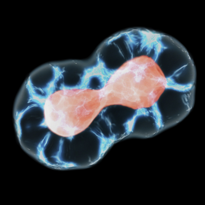
Meiosis is a complex cell-division process that is part of the sexual reproduction cycle in animal, human and plant cells. The end result of meiosis is four haploid daughter cells with half the amount of chromosomes that were in the parent cell before division. Meiosis is broken down into two parts, meiosis I and meiosis II, as the parent cells goes through the process of division twice to make four daughter cells. This differs from mitosis, in which two identical daughter cells are produced.
Cell Structure and Functions of Each Component
Cell Structure
and Functions of Each Component
Eukaryotic cells contain a true nucleus and include cells in humans, animals, plants, fungi and algae that reproduce sexually.
The very exterior of a cell is the cell membrane. This is a semi-permeable barrier that allows only a small number of molecules to move back and forth through it. The cell membrane has a double layer to separate the inner parts of a cell from the outside, but it also allows transport of different substances between the cell and surrounding cells.
Cytoplasm is a fluid that is held inside the cell by the cell membrane. Its job is to support all of the cell structure and shape as well as support the organelles or tiny organs that have specific functions for normal cellular operation.
The nucleus is often called the brain center of the cell. It contains the genetic material or DNA and RNA. It has a nuclear membrane surrounding it with pores to enable protein movement both into and out of it. The nucleolus is inside of the nucleus, and it holds the ribosomes for a cell.
Ribosomes synthesize protein for normal cell functioning. They can be suspended in the cytoplasm or they may be attached to the endoplasmic reticulum. The endoplasmic reticulum is basically the transportation department of a cell and is the means by which proteins move about.
Lysosomes contain digestive enzymes to help break down any waste and remove it from the cell. Lysosomes have a circular shape.
Centrosomes are located near the nucleus of a cell. The centrosome makes microtubules, which aid in cell division of tissues in mitosis by moving the chromosomes to opposite poles of the cell.
Vacuoles are contained by a membrane and are small organelles that store substances and help transport waste out of a cell.
Golgi bodies are also called the Golgi apparatus or the Golgi complex. They form an organelle that packs substances in preparation for transport out of a cell.
Mitochondria are the energy sources of cells. They have a double membrane and take the shape of a sphere or rod. They are located in the cell's cytoplasm, and their function is to convert nutrients and oxygen into energy sources for the cell.
The cell's cytoskeleton helps maintain its shape, using microtubules and fibers. Cilia and flagella are hair-like structures that are present on the cell membrane. These two types of appendages help the cells to move from one place to another.
What Is Meiosis?
What Is Meiosis?
Meiosis is the cell division process for those cells involved in sexual reproduction. A diploid parent cell, which has two complete sets of chromosomes (22 pairs of numbered chromosomes and one pair of sex chromosomes), divides twice to produce four daughter cells that are haploid and each contain half the DNA of the original parent cell before cell division. Meiosis is split into two distinct cycles, I and II, each with its own phases or stages of cell division. Each cycle contains phases, as in mitosis, and each phase is labeled with a number to indicate which cycle it belongs to. For example, meiosis I has prophase I and anaphase I, while meiosis II has prophase II and anaphase II.
What Are the Phases in Meiosis I?
What Are the Phases in
Meiosis I?
Meiosis I, the first half of the total cell division process of sexual reproductive cells, has four phases: prophase I, metaphase I, anaphase I and telophase I. Before mitosis or meiosis I begin, all cells go through interphase.
In interphase , the cell is preparing for cell division and has many functions at this point. The parent cell remains in this phase or stage for most of its life in preparation for division. It is broken down into three smaller subphases: G1 phase, S phase and G2 phase. In the G1 subphase, the parent cell increases in mass so it can later divide into two cells. The G represents the word gap, and the 1 represents the first gap in interphase. The S subphase is next, in which the DNA is synthesized in the parent cell. DNA is replicated in order to provide the two daughter cells in meiosis I with chromosomes from the parent cell. The S stands for synthesis. The next subphase in interphase I is the G2 phase or the second gap phase. In this subphase, the cell increases in size and synthesizes its proteins. The parent cell still has nucleoli present and is bound by the nuclear envelope. The chromosomes are synthesized, but they all remain in the form of chromatin. Centrioles are replicated are located outside of the nucleus.
Prophase I occurs next. The chromosomes in the parent cell start to condense and then attach to the nuclear envelope as synapsis occurs, meaning that a pair of identical chromosomes line up next to each other to form a tetrad. A tetrad is formed from four chromatids. This is the point of genetic recombination or "crossing over" of the genes. Genes are recombined to form new combinations that may or may not be the exact genetic combinations of one parent or another. The chromosomes will then thicken and detach themselves from the nuclear envelope as the centrioles start to move away from each other and the nucleoli and nuclear envelope both break down. The chromosomes will then start their migration to the metaphase plate in anticipation of cell division.
Metaphase I is the next phase in meiosis I. In this phase, the tetrads align themselves at the metaphase plate in the cell, and the centromeres of the chromosome pairs are turned toward the opposite poles or ends of the cell.
Anaphase I is characterized by the chromosomes moving to the opposite sides or poles of the cell. Kinetochore fibers, which are microtubules, start to pull the chromosomes to the opposite cell poles. The sister chromatids remain together after the movement of the chromosomes to opposite poles.
Telophase I is the next phase in meiosis I and also the last phase in this part of meiosis. The spindle fibers continue to pull the chromosome pairs to the opposite poles of the parent cell. After they reach the opposite poles, each pole contains haploid chromosomes, meaning that they each have half the number of chromosomes as the parent cell. The cell divides through cytokinesis in the division of the cytoplasm to produce two daughter haploid cells. Note that at the end of meiosis I, the genetic material does not replicate again.
What Are the Phases of Meiosis II?
What Are the Phases of
Meiosis II?
Meiosis II has four stages, which are prophase II, metaphase II, anaphase II and telophase II.
Metaphase II is characterized when the chromosomes line up at the metaphase II plate in the center of the cell. Note that the metaphase plate from meiosis I is now called the metaphase II plate. The kinetochore fibers of the sister chromatids start to point to opposite sides or poles of the cell.
Anaphase II of meiosis II is the next phase to occur. In it, the sister chromatids separate from each other and start their journey to opposite poles or sides of the cell. At this time, the spindle fibers that are not connected to the chromatids start to lengthen. This causes the cell to elongate its shape. When the pair of sister chromatids separate from each other, they actually become a full chromosome, called daughter chromosomes. The poles of the cell move farther apart as the cell elongates, and at the end of this phase, each pole contains a full set of chromosomes.
Telophase II is the last distinct phase of meiosis II. Nuclei form with one at each opposite pole. Cytokinesis occurs again to divide the cytoplasm and create two more cells. This results in four daughter haploid cells, each containing half the chromosomes as the original parent cell. When sex cells of sperm and eggs unite in fertilization, each pair of joined haploid cells becomes a diploid cell, just as the parent cell was before it began the division process of meiosis.
How Is Meiosis Different From Mitosis?
How Is Meiosis Different From
Mitosis?
All organisms have cells that grow and divide to replace dying cells and to promote growth of the entire organism. This is accomplished through one of two cell division procedures called meiosis and mitosis. Meiosis is the cell division of sexual reproductive cells for gamete formation, and mitosis is the cell division that occurs in all other cells in eukaryotic organisms. Mitosis happens much more often as it includes all body tissues, organs and even hair. Both processes of division are quite similar; however, there are some distinct differences between the two. The differences include the number of daughter cells, the genetic composition, the length of prophase, the formation of tetrads, the chromosome alignment in metaphase and the method of chromosome separation.
In mitosis, a somatic cell that is not a sexual reproduction cell divides only one time. The end product is two daughter cells that are identical at the end of telophase, the last portion of mitosis outside of cytokinesis. In meiosis, a reproductive cell divides once in meiosis I in telophase I and again in meiosis II in telophase II, producing four haploid daughter cells.
The final number of daughter cells produced differs in the two cell division processes with two diploid daughter cells in mitosis and four haploid daughter cells in meiosis.
The genetic composition of the resulting daughter cells also differs between mitosis and meiosis. In mitosis, the two daughter cells are identical. In meiosis, the daughter cells have different genetic combinations due to the process of crossing over.
The length of prophase in mitosis is shorter than the length of prophase I in meiosis; in meiosis, in prophase I, tetrads form with the four chromatids being two sets of sister chromatids; this does not occur in mitosis.
In mitosis, sister chromatids align at the metaphase plate, but in meiosis it is tetrads that align at the metaphase plate in metaphase I.
Sister chromatids separate during anaphase in mitosis to begin migrating toward the opposite poles of a cell. In meiosis, the sister chromatids do not separate from each other in anaphase I.
Cite This Article
MLA
Lougee, Mary. "Meiosis: Definition, Phases 1 & 2, Difference From Mitosis" sciencing.com, https://www.sciencing.com/what-is-meiosis-13714447/. 24 September 2018.
APA
Lougee, Mary. (2018, September 24). Meiosis: Definition, Phases 1 & 2, Difference From Mitosis. sciencing.com. Retrieved from https://www.sciencing.com/what-is-meiosis-13714447/
Chicago
Lougee, Mary. Meiosis: Definition, Phases 1 & 2, Difference From Mitosis last modified August 30, 2022. https://www.sciencing.com/what-is-meiosis-13714447/
