Centriole: Definition, Function & Structure
If you survived biology class, you may recall looking at grainy photos of different cell structures, such as mitochondria, nuclei, or centrioles. In cell biology, a centriole is an organelle, usually near the center of a cell. It plays an important part in cell division, and they are often in pairs located near the nucleus. Importantly, centrioles are only present in eukaryotes (organisms with eukaryotic cells), and they are not found in prokaryotic cells.
Centriole Structure
Centriole Structure
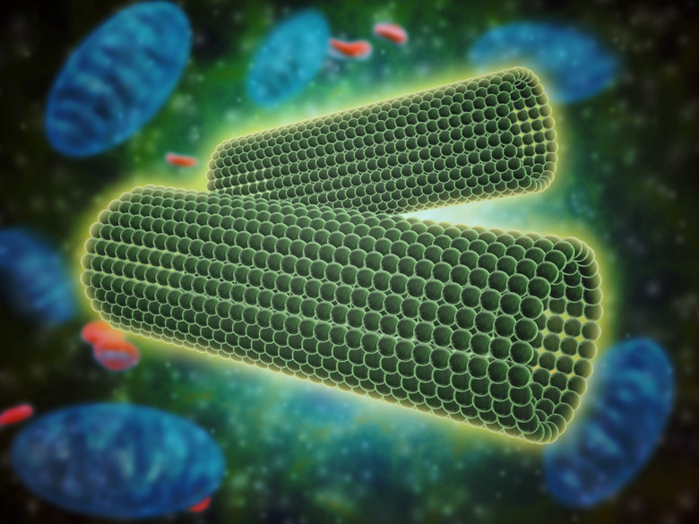
The centrosome contains the centrioles in the cell. Also known as the microtubule-organizing center, the centrosome is an organelle contained within the cytoplasm of the cell. There is typically a pair of centrioles, each with nine bundles of microtubules – which are hollow tubes that give organelles their shape – arranged in a ring. However, some species have fewer than nine bundles. The microtubules run parallel to each other, and each bundle has a set of three microtubules, which are made from a protein called tubulin.
TL;DR (Too Long; Didn't Read)
The main structure of the centriole containing the nine bundles is called a cartwheel. The bundles of microtubule triplets from a very strong structure to support the centriole because of their triple reinforcement.
The two centrioles tend to be oriented at right angles to each other, and they can be classified as a mother and daughter centriole. A centriole looks like a small, hollow cylinder. Unfortunately, you cannot see it clearly until the cell is ready to start division.
In addition to the centrioles, the centrosome contains pericentriolar material (PCM). This is a mass of proteins, which surrounds the two centrioles. Researchers believe that centrioles are capable of organizing the proteins.
Function of Centrioles
Function of Centrioles
The main function of a centriole is to help chromosomes move inside the cell. The centrioles' location depends on whether or not the cell is going through division. You can find centrioles being active during mitosis and meiosis. Mitosis is cell division that leads to two daughter cells with the same number of chromosomes as the original parent cell. On the other hand, meiosis is cell division that leads to daughter cells with half the number of chromosomes as the original parent cell.
When a cell is ready to divide, centrioles move to the opposite ends. During the interphase process of cell division, centrioles control a spindle fiber formation called the mitotic spindle or spindle apparatus. It looks like groups of thread coming out of the centrioles. The spindle is able to pull apart the chromosomes and separate them.
Cell Division Details
Cell Division Details
The centrioles are only active in specific phases of cell division. During prophase of mitosis, the centrosome separates, so a pair of centrioles can travel to opposite sides of the cell. Centriole duplication takes place, and the the centrioles are surrounded with an amorphous pericentriolar material. The centrioles make microtubules, which look like threads and are called spindle fibers.
The microtubules start growing toward the opposite end of the cell. Then, some of these microtubules attach to the centromeres of chromosomes. A portion of the microtubules will help separate the chromosomes, while the others will help the cell split into two. Eventually, the chromosomes line up in the middle of the cell. This is called metaphase.
Next, during anaphase, the sister chromatids begin to separate, and the halves move along the microtubule threads. During telophase, the chromatids move to opposite ends of the cell. At this time, the centrioles' spindle fibers start to disappear since they are not needed. After the centrioles serve their role, replication of chromosomes takes place, and the process of cytokinesis (cell division) can take place.
Centromeres and Cytoskeletons
Centromeres and Cytoskeletons
A centromere is a region on a chromosome that allows attachment from the microtubules from the centriole. When you look at a picture of a chromosome, the centromere appears as the constricted area in the middle. In this region, you can find specialized chromatin. Centromeres play an essential part in the separation of chromatids during cell division. It is important to note that although most biology textbooks show the centromere in the middle of the chromosome, the position can vary. Some centromeres are in the middle, while others are closer to the ends. Centromeres and centrioles are quite distinct, but they both participate in the interphase process of genome duplication.
The cytoskeleton creates an internal structure to help support the cell; it consists of microtubules and other proteins (distinct from centriolar microtubules). The cytoskeleton tubules can be thought as spokes positioned to support the cell from the inside.
Cilia and Flagella
Cilia and Flagella
You can also see centrioles at the basal ends of flagella and cilia, which are projections coming out of a cell (outside of the cell membrane); this is why they are sometimes called basal bodies. The microtubules in centrioles make up the flagellum or cilium, which are designed to either help the cell move or to help it control substances around it.
When centrioles move to the periphery of a cell, they can organize and form the cilia and flagella. Cilia tend to be composed of many small projections, and they may look like little hairs covering a cell. Some examples of cilia are the projections on the tissue surface of a mammal's trachea. On the other hand, flagella are different and only have one long projection. It often looks like a tail. One example of a cell with a flagellum is a mammalian sperm cell.
Most eukaryotic cilia and flagella have similar internal structures made up of microtubules. They are called doublet microtubules and are arranged in a nine plus two fashion. Nine doublet microtubules, consisting of two pieces, circle two inner microtubules.
TL;DR (Too Long; Didn't Read)
It is rare, but single microtubules can be found in the embryo cells of caenorhabditis elegans.
Cells That Have Centrioles
Cells That Have Centrioles
Only animal cells have centrioles, so bacteria, fungi and algae do not have them. Some lower plant cells have centrioles, but higher plants do not. Generally, lower plants include mosses, lichens and liverworts because they do not have a vascular system. On the other hand, higher plants have this system and include shrubs, trees and flowers.
The exception are ciliated cells that may not have internal centriole organelles, but they have the structure of centriole microtubules outside their membrane.
Centrioles and Diseases
Centrioles and Diseases
When mutations occur in the genes that are responsible for the proteins found in centrioles, problems and genetic diseases can happen. Scientists think that centrioles may actually carry biological information. It is important to note that in a fertilized egg the centrioles only come from the male's sperm because a female's egg does not contain them. Researchers have found that the original centrioles from the sperm are able to survive multiple cell divisions in the embryo.
Although centrioles do not carry genetic information, their persistence in a developing embryo means they can contribute other types of information. The reason why scientists are interested in this topic is the potential it holds for understanding and treating diseases associated with centrioles. For example, centrioles that have problems in a male's sperm can be passed on to the embryo.
Centrioles and Cancer
Centrioles and Cancer
Researchers have discovered that cancer cells often have more centrioles than necessary. Not only do they have extra centrioles, but they also have longer ones than normal. However, when scientists removed centrioles from cancer cells in a study, they found that the cells could continue to divide at a slower rate. They learned that cancer cells have a mutation in p53, which is a gene that codes for a protein responsible for controlling the cell cycle, so they can divide at an accelerated rate. Scientists believe this discovery will help improve cancer treatments.
Oral-Facial-Digital (OFD) Syndrome
Oral-Facial-Digital (OFD) Syndrome
Oral-facial-digital (OFD) syndrome is a genetic disorder that is also abbreviated as OFDS. This congenital disease happens because of problems with cilia that lead to signaling issues. Researchers found that mutations in two genes, OFD1 and C2CD3, can lead to problems with proteins in centrioles. Both of these genes are responsible for regulating centrioles, but mutations prevent the proteins from functioning normally. This leads to defective cilia.
Oral-facial-digital syndrome causes developmental abnormalities in humans. It affects the head, mouth, jaw, teeth and other parts of the body. In general, people with this condition have issues with the oral cavity, their face and digits. OFDS can also lead to intellectual disabilities. There are different types of oral-facial-digital syndrome, but some are hard to distinguish from each other.
Some of the OFDS symptoms include cleft palate, cleft lip, small jaw, hair loss, tongue tumors, small or wideset eyes, extra digits, seizures, growth problems, heart and kidney disease, sunken chest and skin lesions. It is also common for people with OFDS to have extra or missing teeth. An estimated one in 50,000 to 250,000 births results in oral-facial-digital syndrome. OFD syndrome type I is the most common of all the types.
A genetic test can confirm oral-facial-digital syndrome because it can show the gene mutations that cause it. Unfortunately, it only works for diagnosing OFD syndrome type I and not the other types. The others are usually diagnosed based on symptoms. There is no cure for OFDS, but plastic or reconstructive surgery may help correct some of the facial abnormalities.
Oral-facial-digital syndrome is an X-linked genetic disorder. This means a mutation occurs on the X chromosome, which is inherited. When a female has a mutation on at least one X chromosome out of two, she will have the disorder. However, since males only have one X chromosome, if they get a mutation, it tends to be lethal. This results in more females than males having OFDS.
Meckel-Gruber Syndrome
Meckel-Gruber Syndrome
Meckel-Gruber syndrome, which is also called Meckel syndrome or Gruber syndrome, is a genetic disorder. It is also caused by defects in cilia. Meckel-Gruber syndrome affects different organs in the body, including the kidneys, brain, digits and liver. The most common symptoms are having a protrusion on part of the brain, kidney cysts, and extra digits.
Some people with this genetic disease have facial and head abnormalities. Others have brain and spinal cord problems. Many fetuses that have Meckel-Gruber syndrome die before birth, and those who are born tend to live for a short time. They often suffer respiratory or kidney failure.
An estimated one in 3,250 to 140,000 babies has this genetic disorder. However, it is more common in certain parts of the world and some countries. For example, it occurs in one in 9,000 people with Finnish ancestry, one in 3,000 people with Belgian ancestry and one in 1,300 people with Gujarati Indian ancestry.
Most fetuses are diagnosed during pregnancy, when an ultrasound is performed. It can show a brain abnormality that looks like a protrusion. Pregnant women may also get chorionic villus sampling or amniocentesis to check for the disorder. A genetic test can also confirm diagnosis. There is no cure for Meckel-Gruber syndrome.
Mutations in multiple genes can lead to Meckel-Gruber syndrome. This creates proteins that cannot function properly, and the cilia are affected negatively. The cilia have both structural and functional issues, which cause signaling abnormalities inside cells. Meckel-Gruber syndrome is an autosomal recessive condition. This means there are mutations on both copies of a gene that a fetus inherits.
Centriole Importance
Centriole Importance
Centrioles are important organelles inside cells. They are part of cell division, cilia and flagella. However, when problems occur, it can lead to several diseases. When a mutation in a gene causes protein malfunctions that affect cilia, it can lead to serious genetic disorders that are fatal. Researchers continue to study centrioles to learn more about their function and structure, with the hope of saving and improving lives in the future.
References
- Molecular Expressions Cell Biology: Animal Cell Structure – Centrioles
- BMC Biology: Q&A Who needs a centrosome?
- ScienceDirect: Centriole
- Phys.org: Beyond genes: Are centrioles carriers of biological information?
- ASCB: Over 100 Years Later, Centriole Debate Settled with Potential Cancer Therapeutic
- NORD: Oral-Facial-Digital Syndrome
- GHR: Meckel syndrome
Cite This Article
MLA
Bandoim, Lana. "Centriole: Definition, Function & Structure" sciencing.com, https://www.sciencing.com/what-is-the-centriole-13714443/. 12 July 2023.
APA
Bandoim, Lana. (2023, July 12). Centriole: Definition, Function & Structure. sciencing.com. Retrieved from https://www.sciencing.com/what-is-the-centriole-13714443/
Chicago
Bandoim, Lana. Centriole: Definition, Function & Structure last modified July 12, 2023. https://www.sciencing.com/what-is-the-centriole-13714443/