The Advantages Of Stained Bacteria
Microbiologists study the characteristics of microorganisms such as algae, protozoa, bacteria, fungi and viruses using a microscope. While some organisms such as protozoa and yeast cells are easy to observe using a wet mount, bacterial cells require staining. Scientists developed several methods such as Gram staining, acid-fast staining and fluorescent staining for better visualization of bacterial cells and cellular structures. Using such staining methods, it is possible to identify structural features that help classify bacteria.
Better Visualization
Better Visualization
Bacterial organisms are so small that most of them are visible only under a microscope with a magnification power of 1000X. However, mere magnification of size does not provide a sufficient degree of clarity, so that bacteria must therefore be stained before observation to provide the clarity needed for visualization.
Identification and Classification
Identification and Classification
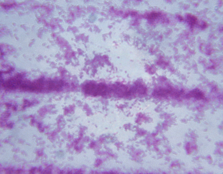
Staining bacteria to distinguish between bacterial types is known as differential staining. The Gram stain is one such differential stain that distinguishes between bacteria on the basis of their cell wall content. In this method, the bacterial cells react with a crystal violet stain to take up a violet color. On adding a de-staining agent, some bacterial cells lose color whereas others don't. On adding safranin stain, the decolorized cells take up the stain to appear red while the bacterial cells that didn't lose color remain violet. The bacterial cells that take up the red color are called Gram negative organisms and those that don't take up the color are classified as Gram positive organisms. Gram staining provides a rapid method for the initial identification of bacteria involved in infections. Similarly, the acid-fast staining procedure helps to specifically identify the organisms belonging to the class of bacteria called Mycobacteria, such as Mycobacterium tuberculosis.
Detection of Viability
Detection of Viability
In bacterial culture specimens, it is often important to detect the presence of living bacterial cells. Staining methods such as fluorescent staining help to identify if culture cells are viable or not. Living bacteria have the ability to convert the 5-Cyano-2,3-ditolyl Tetrazolium Chloride (CTC) stain into a dye which shows a red fluorescence. Therefore, when cultures stained with CTC emit such fluorescence, it indicates the presence of viable bacteria. Propidium iodide is a stain that acts only on non-living cells that possess damaged membranes, and therefore, is useful in identifying dead bacterial cells.
Identification of Cellular Structures
Identification of Cellular Structures
Staining provides a method of clearly visualizing several cellular structures. For example, the Fuelgen staining method allows identification of the nucleus within bacterial cells whereas Albert's stain is useful in visualizing metachromatic granules. Similarly, the silver impregnation technique allows identification of spirochetes. Flagella are easy to observe when stained with Ryu's stain. Malachite green staining helps to identify bacterial spores.
Cite This Article
MLA
Stewart, David. "The Advantages Of Stained Bacteria" sciencing.com, https://www.sciencing.com/advantages-stained-bacteria-12011291/. 24 April 2017.
APA
Stewart, David. (2017, April 24). The Advantages Of Stained Bacteria. sciencing.com. Retrieved from https://www.sciencing.com/advantages-stained-bacteria-12011291/
Chicago
Stewart, David. The Advantages Of Stained Bacteria last modified March 24, 2022. https://www.sciencing.com/advantages-stained-bacteria-12011291/