Cell Cycle: Definition, Phases, Regulation & Facts
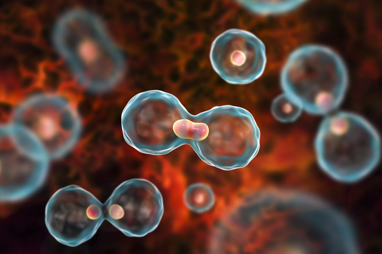
Cell division is vital to the growth and health of an organism. Almost all cells engage in cell division; some do it multiple times in their lifespans. A growing organism, such as a human embryo, uses cell division to increase the size and specialization of individual organs. Even mature organisms, like a retired adult human, use cell division to maintain and repair body tissue. The cell cycle describes the process by which cells do their designated jobs, grow and divide, and then begin the process again with the two resulting daughter cells. In the 19th century, technological advances in microscopy allowed scientists to determine that all cells arise from other cells through the process of cell division. This finally disproved the previously widespread belief that cells generated spontaneously from available matter. The cell cycle is responsible for all ongoing life. Regardless of whether it happens in the cells of algae clinging to a rock in a cave or in the cells of the skin on your arm, the steps are the same.
TL;DR (Too Long; Didn't Read)
Cell division is vital to the growth and health of an organism. The cell cycle is the repeating rhythm of cell growth and division. It consists of the stages interphase and mitosis, as well as their subphases, and the process of cytokinesis. The cell cycle is strictly regulated by chemicals at checkpoints throughout each step to make sure that mutations do not occur and that cell growth does not happen faster than what is healthy for the surrounding tissue.
The Phases of the Cell Cycle
The Phases of the Cell Cycle
The cell cycle essentially consists of two phases. The first phase is interphase. During interphase, the cell is preparing for cell division in three subphases called G1 phase, S phase and G2 phase. By the end of interphase, the chromosomes in the cell nucleus have all been duplicated. Through all of these stages, the cell is also continuing to go about its daily functions, whatever those are. Interphase can last days, weeks, years – and in some cases, for the entire lifespan of the organism. Most nerve cells never leave the G1 stage of interphase, so scientists have designated a special stage for cells like them called G0. This stage is for nerve cells and other cells that will not be going into a process of cell division. Sometimes this is because they are simply not ready to or not designated to, like nerve cells or muscle cells, and that is called a state of quiescence. Other times, they are too old or damaged, and that is called a state of senescence. Since nerve cells are separate from the cell cycle, damage to them is mostly irreparable, unlike a broken bone, and this is the reason that people with spine or brain injuries often have permanent disabilities.
The second phase of the cell cycle is called mitosis, or M phase. During mitosis, the nucleus divides in two, sending one copy of each duplicated chromosome to each of the two nuclei. There are four stages of mitosis, and these are prophase, metaphase, anaphase and telophase. At approximately the same time that mitosis is happening, another process occurs, called cytokinesis, which is almost its own phase. This is the process by which the cell's cytoplasm, and everything else in it, divides. That way, when the nucleus splits in two, there are two of everything in the surrounding cell to go with each nucleus. Once the dividing is complete, the plasma membrane closes around each new cell and pinches off, dividing the two new identical cells from each other completely. Immediately, both cells are in the first stage of interphase again: G1.
Interphase and Its Subphases
Interphase and Its Subphases
G1 stands for Gap phase 1. The term "gap" comes from a time when scientists were discovering cell division under microscope and found the mitotic stage very exciting and important. They observed the nucleus dividing and the accompanying cytokinetic process as proof that all cells came from other cells. The stages of interphase, however, seemed static and inactive. Therefore, they thought of them as resting periods, or gaps in activity. The truth, however, is that G1 – and G2 at the end of interphase – are bustling growth periods for the cell, in which the cell grows in size and contributes to the well-being of the organism in whatever way it was "born" to do. In addition to its regular cellular duties, the cell builds molecules such as proteins and ribonucleic acid (RNA).
If the cell's DNA is not damaged and the cell has grown enough, it proceeds into the second stage of interphase, called S phase. This is short for Synthesis phase. During this phase, as the name suggests, the cell devotes a good deal of energy to synthesizing molecules. Specifically, the cell replicates its DNA, duplicating its chromosomes. Humans have 46 chromosomes in their somatic cells, which are all cells that aren't reproductive cells (sperm and ova). The 46 chromosomes are organized into 23 homologous pairs that are joined together. Each chromosome in a homologous pair is called the other one's homolog. When the chromosomes are duplicated during S phase, they are coiled very tightly around histone protein strands called chromatin, which makes the duplication process less prone to DNA replication errors, or mutation. The two new identical chromosomes are now each called chromatids. Strands of histones bind the two identical chromatids together so that they form a kind of X shape. The point where they are bound is called a centromere. In addition, the chromatids are still joined to their homolog, which is now also an X-shaped pair of chromatids. Each pair of chromatids is called a chromosome; the rule of thumb is that there is never more than one chromosome attached to one centromere.
The last stage of interphase is G2, or Gap phase 2. This phase was given its name for the same reasons as G1. Just like during G1 and S phase, the cell remains busy with its typical tasks throughout the stage, even as it finishes the work of interphase and prepares for mitosis. To prepare for mitosis, the cell divides its mitochondria, as well as its chloroplasts (if it has any). It begins to synthesize the precursors of spindle fibers, which are called microtubules. It makes these by replicating and stacking the centromeres of the chromatid pairs in its nucleus. Spindle fibers will be crucial to the process of nuclear division during mitosis, when chromosomes will have to be pulled apart into the two separating nuclei; making certain that the correct chromosomes get to the correct nucleus and remain paired with the correct homolog are crucial, to prevent genetic mutations.
The Breakdown of the Nuclear Membrane in Prophase
The Breakdown of the Nuclear Membrane in Prophase
The dividing markers between the phases of the cell cycle and the subphases of interphase and mitosis are artifices that scientists use to be able to describe the process of cell division. In nature, the process is fluid and never-ending. The first stage of mitosis is called prophase. It begins with the chromosomes in the state they were in at the end of the G2 stage of interphase, replicated with sister chromatids attached by centromeres. During prophase, the chromatin strand condenses, which allows the chromosomes (that is, each pair of sister chromatids) to become visible under light microscopy. The centromeres continue to grow into microtubules, which form spindle fibers. By the end of prophase, the nuclear membrane breaks down, and the spindle fibers connect to form a structural network throughout the cytoplasm of the cell. Since the chromosomes are now floating free in the cytoplasm, the spindle fibers are the only support that keeps them from floating astray.
The Spindle Equator in Metaphase
The Spindle Equator in Metaphase
The cell moves into metaphase as soon as the nuclear membrane dissolves. The spindle fibers move the chromosomes to the cell's equator. This plane is known as the spindle equator or the metaphase plate. There is nothing tangible there; it is simply a plane where all of the chromosomes line up, and which bisects the cell either horizontally or vertically, depending on how you are viewing or imagining the cell (for a visual representation of this, see Resources). In humans, there are 46 centromeres, and each one is attached to a pair of chromatid sisters. The number of centromeres depends on the organism. Each centromere is connected to two spindle fibers. The two spindle fibers diverge once they leave the centromere, so that they connect to structures on opposite poles of the cell.
Two Nuclei in Anaphase and Telophase
Two Nuclei in Anaphase and Telophase
The cell shifts into anaphase, which is the briefest of the four phases of mitosis. The spindle fibers that connect the chromosomes to the poles of the cell shorten and move away toward their respective poles. In doing so, they pull apart the chromosomes they are attached to. The centromeres also split in two as one half travels with each chromatid sister toward an opposite pole. Since each chromatid now has its own centromere, it is called a chromosome again. Meanwhile, different spindle fibers attached to both poles lengthen, causing the distance between the two poles of the cell to grow, so that the cell flattens and elongates. The process of anaphase happens in such a way so that by the end, each side of the cell contains one copy of each chromosome.
Telophase is the fourth and final stage of mitosis. In this stage, the extremely tightly packed chromosomes – which were condensed to increase accuracy of replication – uncoil themselves. The spindle fibers dissolve, and a cellular organelle called the endoplasmic reticulum synthesizes new nuclear membranes around each set of chromosomes. This means that the cell now has two nuclei, each with a complete genome. Mitosis is complete.
Animal and Plant Cytokinesis
Animal and Plant Cytokinesis
Now that the nucleus has been divided, the rest of the cell needs to divide as well so that the two cells can part. This process is known as cytokinesis. It is a separate process from mitosis, although it often co-occurs with mitosis. It happens differently in animal and plant cells, because where animal cells only have a plasma cell membrane, plant cells have a rigid cell wall. In both kinds of cells, there are now two distinct nuclei in one cell. In animal cells, a contractile ring forms at the midpoint of the cell. This is a ring of microfilaments that cinch around the cell, tightening the plasma membrane at the center like a corset until it creates what is known as a cleavage furrow. In other words, the contractile ring causes the cell to form an hourglass shape that becomes more and more pronounced, until the cell pinches off into two separate cells entirely. In plant cells, an organelle called the Golgi complex creates vesicles, which are membrane-bound pockets of liquid along the axis that divides the cell between the two nuclei. Those vesicles contain polysaccharides that are needed to form the cell plate, and the cell plate eventually fuses with and becomes part of the cell wall that once housed the original single cell, but is now home to two cells.
Cell Cycle Regulation
Cell Cycle Regulation
The cell cycle requires a great deal of regulation to make sure that it does not proceed without certain conditions being met inside and outside of the cell. Without that regulation, there would be an unchecked genetic mutations, out-of-control cell growth (cancer), and other problems. The cell cycle has a number of checkpoints to make sure that things are proceeding correctly. If they are not, repairs are made, or programmed cell death is initiated. One of the primary chemical regulators of the cell cycle is cyclin-dependent kinase (CDK). There are different forms of this molecule that operate at different points in the cell cycle. For example, the protein p53 is produced by damaged DNA in the cell, and which will deactivate the CDK complex at the G1/S checkpoint, thus arresting the cell's progress.
Cite This Article
MLA
E., Rebecca. "Cell Cycle: Definition, Phases, Regulation & Facts" sciencing.com, https://www.sciencing.com/cell-cycle-20206/. 23 April 2019.
APA
E., Rebecca. (2019, April 23). Cell Cycle: Definition, Phases, Regulation & Facts. sciencing.com. Retrieved from https://www.sciencing.com/cell-cycle-20206/
Chicago
E., Rebecca. Cell Cycle: Definition, Phases, Regulation & Facts last modified August 30, 2022. https://www.sciencing.com/cell-cycle-20206/
