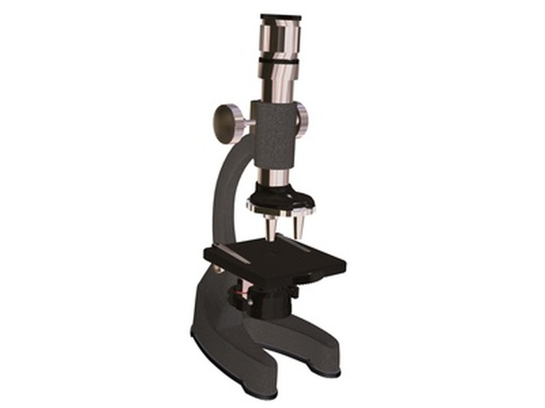
How To Make Histology Slides
Histology is the microscopic study of tissues. It is a natural extension of microscopic cell biology. Rather than simply examining different kinds of cells in isolation, microbiologists more often look at groups of adjacent cells that form common kinds of tissue. In humans, there are four basic types of tissues made from constituent cells that also have significantly different properties.
Medical microbiologists, who work in tandem with physicians and other clinicians, often must prepare and examine tissues quickly so that important decisions can be made, such as whether a patient already in surgery should have an organ removed thanks to suspected cancer. The histology professionals can often determine this on the basis of the microscopic appearance of cells and tissues.
The Importance of Microbiology
The Importance of Microbiology
Microbiology's importance in various medical realms cannot be overstated, but this is perhaps most evident in the fighting of infectious disease. Virtually all pathogens (disease-causing life forms) are too small to be seen with the unaided eye.
Without microbiology, not only would humans not know for sure what even causes infectious diseases, but researchers would also be unable to distinguish between different types of disease-causing organisms, leaving the species effectively defenseless against bacterial, viral, fungal and protozoan invaders.
The Early History of Microscopy
The Early History of Microscopy
The first known compound microscope – that is, a microscope that uses more than one lens to create an image – entered the technology scene in 1590. While multiple inventors were simultaneously at work on such a device, its invention is usually credited to the father-and-son team of Hans and Zacharias Jensen.
Oddly, it was not until the 1660s or so that people started to consider the potential for using what by then had come to be called "microscopes" to look at very tiny things. Up to that point, scientists had been more interested in making very small but visible things look very large and detailed. Soon afterward, Antony Van Leeuwenhoek discovered bacteria.
The Four Basic Tissue Types
The Four Basic Tissue Types
Anatomists have divided human tissue into four types: epithelium, connective tissue, nervous tissue and muscle. Each of these has numerous subtypes revealed by close microscopic examination. Different tissue types tend to come from different layers formed early in the embryo stage of development in the womb.
Epithelial tissue lines the body's hollow organs and also the outer surface of the body, in the form of skin. Connective tissue includes cartilage, bone, blood cells and adipose (fat) tissue, and includes both loose and dense types.
Nervous tissue makes up the brain, spinal cord and peripheral nerves, and includes neurons (nerve cells) and glial cells (support cells of neurons). Muscle tissue makes up skeletal muscles (your more obvious muscles), the smooth muscles of internal organs and the muscle of the heart.
Making Histology Slides
Making Histology Slides
To make your own histology slides, you'll need more than what you are likely to find sitting around the house. Making histology slides is more than a simple matter of swabbing samples onto pieces of glass.
For example, some histological sections need to be quite thin, and so any university histology lab is likely to feature a vibratome, which is essentially a tiny knife for making histology "slices" to be viewed under a microscope.
Tissue protectors and preservers, automatic stainers and cryostats (for working with frozen stainers) are some of the other things you're likely to see if you glance around a histology lab and look closely at the labels of the equipment.
Histology sample preparation and general histology steps vary widely from lab to lab and depend, naturally, on the nature of the sample and the purpose for examining it. If you are taking part in any experiments, make sure you know the rules of the room, especially the safety rules.
Cite This Article
MLA
Beck, Kevin. "How To Make Histology Slides" sciencing.com, https://www.sciencing.com/make-histology-slides-7348900/. 14 June 2019.
APA
Beck, Kevin. (2019, June 14). How To Make Histology Slides. sciencing.com. Retrieved from https://www.sciencing.com/make-histology-slides-7348900/
Chicago
Beck, Kevin. How To Make Histology Slides last modified August 30, 2022. https://www.sciencing.com/make-histology-slides-7348900/
