Prokaryotic Cell Structure

When you think about cells and cell structure, you probably picture highly organized, organelle-rich eukaryotic cells, such as those that make up your own body. The other type of cell, called a prokaryotic cell, is quite different from what you picture (although no less fascinating).
For one thing, prokaryotic cells are much smaller than eukaryotic cells. Each prokaryote is about one-tenth the size of a eukaryote or about the size of the eukaryotic cell's mitochondria.
Prokaryotic Cell Structure
Prokaryotic Cell Structure
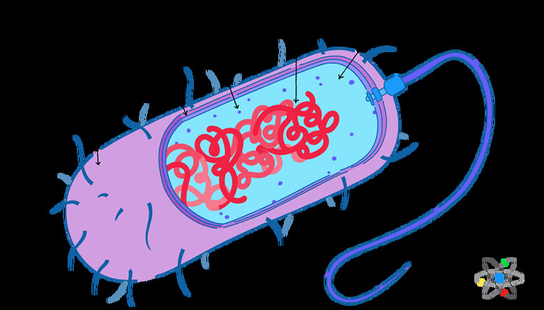
The typical prokaryotic cell is also much simpler than eukaryotic cells when it comes to cell structure and organization. The word prokaryote comes from the Greek words pro, meaning before, and karyon, meaning nut or kernel. For scientists who study prokaryotic cells, this somewhat mysterious language refers to **organelles**, especially the nucleus.
Put simply, prokaryotic cells are unicellular organisms that do not have a nucleus or other membrane-bound organelles like eukaryotic cells do: they lack organelles.
Still, prokaryotes share many underlying characteristics with eukaryotes. While they are smaller and less complex than their eukaryote cousins, prokaryotic cells still have defined cell structures, and learning about those structures is important for understanding unicellular organisms, such as bacteria.
The Nucleoid
The Nucleoid
While prokaryotic cells do not have membrane-bound organelles like a nucleus, they do have a region within the cell dedicated to DNA storage called the nucleoid. This area is a distinct section of the prokaryotic cell but is not walled off from the rest of the cell by a membrane. Instead, the majority of the cell's DNA simply stays near the center of the prokaryotic cell.
This prokaryotic DNA is quite a bit different from eukaryotic DNA, too. It is still tightly coiled and contains the cell's genetic information, but for prokaryotic cells, this DNA exists as one large loop or ring.
Some prokaryotic cells also have additional rings of DNA called **plasmids**. These plasmids do not localize in the center of the cell, only contain a few genes and replicate independently from the chromosomal DNA in the nucleoid.
Ribosomes
Ribosomes
All the area inside the plasma membrane of a prokaryotic cell is the **cytoplasm**. In addition to the nucleoid and plasmids, this space contains a substance called cytosol, which has the consistency of jelly. It also contains ribosomes scattered throughout the cytosol.
These prokaryotic ribosomes are not organelles since they do not have membranes, but they still perform functions similar to those performed by eukaryotic ribosomes. This includes two vital roles:
- Gene expression
- Protein synthesis
You might be surprised to learn just how abundant ribosomes are in prokaryotic cells. For example, one prokaryotic unicellular organism called Escherichia coli, which is a type of bacteria that lives in your intestines, contains about 15,000 ribosomes. That means ribosomes make up approximately a quarter of the mass of the entire E. coli cell.
Those many prokaryotic ribosomes contain protein and RNA and have two parts or subunits. Together, these subunits take the genetic material transcribed from the prokaryotic DNA by specialized RNA messengers and convert the data into strings of amino acids. Once folded, those amino acid chains are functional proteins.
Prokaryote Cell Wall Structure
Prokaryote Cell Wall Structure
One of the most important features of prokaryotic cells is the **cell wall**. While eukaryotic plant cells also contain a cell wall, eukaryotic animal cells do not. This rigid barrier is the outside layer of the cell, which separates the cell from the outside world. You can think of the cell wall as a shell, sort of like the shell covering and protecting an insect.
A cell wall is very important for the prokaryotic cell because it:
- Gives the cell its shape
- Keeps the contents of the cell from leaking out
- Protects the cell from damage
The cell wall gets its structure from carbohydrate chains of simple sugars called polysaccharides.
The specific structure of the cell wall depends on the type of prokaryote. For example, the structural components of archaea cell walls vary a great deal. These are generally made of various polysaccharides and glycoproteins but do not contain peptidoglycans like those found in the cell walls of bacteria.
Bacterial cell walls are usually made of peptidoglycans. These cell walls also vary a bit, depending on the type of bacteria they protect. For example, gram positive bacteria (which turn purple or violet during Gram staining in the lab) have thick cell walls while gram negative bacteria (which turn pink or red during Gram staining) have thinner cell walls.
The crucial nature of cell walls comes into stark focus when you consider the way medicine works and how it affects different types of bacteria. Many antibiotics try to pierce the bacterial cell wall in order to kill the bacteria causing an infection.
A rigid cell wall that is impervious to this attack will help the bacteria survive, which is great news for the bacteria and not great for the infected person or animal.
Cell Capsule
Cell Capsule
Some prokaryotes take cell defense a step further by forming yet another protective layer around the cell wall called a capsule. These structures:
- Help prevent the cell from drying out
- Protect against destruction
For this reason, bacteria with capsules may be more difficult to eradicate naturally by the immune system or medically with antibiotics.
For example, the bacteria Streptococcus pneumoniae, which can cause pneumonia, have a capsule covering its cell wall. Variations of the bacteria that no longer have a capsule do not cause pneumonia since they are easily taken up and destroyed by the immune system.
Cell Membrane
Cell Membrane
One similarity between eukaryotic cells and prokaryotes is that they both have a **plasma membrane**. Just under the cell wall, prokaryotic cells have a cell membrane composed of fatty phospholipids.
This membrane, which is actually a lipid bilayer, contains both proteins and carbohydrates.
These protein and carbohydrate molecules play important roles in the plasma membrane as they help cells communicate with each other and also move cargo into and out of the cell.
Some prokaryotes actually contain two cell membranes instead of one. Gram negative bacteria have a traditional inner membrane, which is between the cell wall and the cytoplasm, and an outer membrane just outside the cell wall.
Pili Projections
Pili Projections
The word pilus (plural is pili) comes from the Latin word for hair.
These hair-like projections stick out from the surface of the prokaryotic cell and are important for many types of bacteria. The pili enable a unicellular organism to interact with other organisms using receptors and help them cling to things to avoid being removed or washed away.
For example, useful bacteria that live in your intestines may use pili to hang onto the epithelial cells lining the walls of your intestines. Less friendly bacteria also take advantage of pili to make you sick. These pathogenic bacteria use pili to hold themselves in place during infection.
Very specialized pili called sex pili make it possible for two bacterial cells to come together and exchange genetic material during sexual reproduction called **conjugation**. Since the pili are very fragile, the turnover rate is high, and prokaryotic cells continually make new ones.
Fimbriae and Flagella
Fimbriae and Flagella
Gram negative bacteria may also have fimbriae, which are thread-like, and help anchor the cell to a substrate. For example, Neisseria gonorrhoeae, the gram negative bacteria that causes gonorrhea, uses fimbriae to stick to the membranes during infection with the sexually transmitted disease.
Some prokaryotic cells use whip-like tails called **flagellum** (plural is flagella) to enable cell movement. This whipping structure is actually a hollow, helix-shaped tube made from a protein called flagellin.
These appendages are important for both gram negative bacteria and gram positive bacteria. However, the presence or absence of flagella may depend on the cell's shape since spherical bacteria, called cocci, usually don't have flagella.
Some rod-shaped bacteria, such as Vibrio cholerae, the microbe that causes cholera, have a single whipping flagellum at one end.
Other rod-shaped bacteria, like Escherichia coli, have many flagella covering the entire cell surface. Flagella may have a rotary motor structure located at the base, which enables the whipping motion and therefore bacterial movement or locomotion. Approximately half of all known bacteria have flagella.
 Sciencing
Nutrient Storage
Nutrient Storage
Prokaryotic cells often live under harsh conditions. Ongoing access to nutrients the cell needs to survive may be unreliable causing times of excess nutrients and times of starvation. To deal with this ebb and flow of nourishment, prokaryotic cells developed structures for nutrient storage.
This enables unicellular organisms to take advantage of times rich in nutrients by storing those things in anticipation of future nutrient shortages. Other storage structures evolved to help prokaryotic cells better produce energy, especially under difficult circumstances like aquatic environments.
One example of an adaptation that enables energy production is the gas vacuole or gas vesicle.
These storage compartments are spindle-shaped, or wider through the midsection and tapered at the ends, and formed by a shell of proteins. These proteins keep water out of the vacuole while allowing gases to enter and exit. Gas vacuoles act like internal flotation devices, decreasing the cell's density when filled with gas in order to make the unicellular organism more buoyant.
Gas Vacuole and Photosynthesis
Gas Vacuole and Photosynthesis
This is especially important for prokaryotes that live in water and need to perform photosynthesis for energy, such as planktonic bacteria.
Thanks to the buoyancy provided by gas vacuoles, these unicellular organisms do not sink too deep into the water where it would be more difficult (or even impossible) to capture the sunlight they need to produce energy.
Storage for Misfolded Proteins
Storage for Misfolded Proteins
Another type of storage compartment holds proteins. These inclusions or inclusion bodies usually contain misfolded proteins or foreign materials. For instance, if a virus infects a prokaryote and replicates inside it, the resultant proteins may not be foldable using the prokaryote's cell components.
The cell simply stores these things in inclusion bodies.
This also sometimes happens when scientists use prokaryotic cells for cloning. For example, scientists produce the insulin that people with diabetes rely on to survive using a bacterial cell with a cloned insulin gene.
Learning how to do this correctly required a lot of trial and error for the researchers since the bacterial cells struggled to process the cloned information, instead forming inclusion bodies filled with foreign proteins.
Specialized Microcompartments
Specialized Microcompartments
Prokaryotes also contain protein microcompartments for other types of specialized storage. For example, prokaryotic unicellular organisms that use photosynthesis to make energy, such as autotrophic bacteria, use carboxysomes.
These storage compartments hold the enzymes the prokaryotes need for carbon fixation. This occurs during the second half of photosynthesis when autotrophs convert carbon dioxide to organic carbon (in the form of sugar) using enzymes stored in carboxysomes.
One of the most interesting types of prokaryotic protein microcompartment is the magnetosome.
These specialized storage units contain 15 to 20 magnetite crystals, each covered with a lipid bilayer. Together, these crystals act like the needle of a compass, giving the prokaryotic bacteria that have them the ability to sense the magnetic field of the Earth.
These prokaryotic single celled organisms use this information to orient themselves.
Related prokaryotic cell topics:
Cite This Article
MLA
Mayer, Melissa. "Prokaryotic Cell Structure" sciencing.com, https://www.sciencing.com/prokaryotic-cell-structure-13717682/. 21 May 2019.
APA
Mayer, Melissa. (2019, May 21). Prokaryotic Cell Structure. sciencing.com. Retrieved from https://www.sciencing.com/prokaryotic-cell-structure-13717682/
Chicago
Mayer, Melissa. Prokaryotic Cell Structure last modified August 30, 2022. https://www.sciencing.com/prokaryotic-cell-structure-13717682/