How To Make A DNA Model Out Of Beads & Straws
Deoxyribonucleic acid (DNA) models began with the X-ray diffraction pictures taken by Rosalind Franklin. Her photographs helped Francis Crick and James Watson complete their three-dimensional model of DNA, the now-famous double helix.
While models of DNA can be purchased, building a model helps understand the structure.
DNA Double Helix Model
DNA Double Helix Model
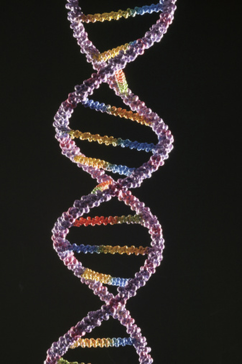
The DNA double helix model contains six parts. The backbone, or sides, of the model consists of phosphate molecules alternating with deoxyribose molecules. The nitrogenous bases of the DNA molecule only connect with the deoxyribose molecules, not with the phosphate molecules.
About 60 percent of the rungs of the DNA molecule are made of adenine-thymine nitrogenous bases. About 40 percent of the rungs are made of guanine-cytosine bases. If the model has 10 rungs, six rungs will be adenine-thymine rungs, and the remaining four rungs will be guanine-cytosine rungs.
Adenine and thymine connect with two hydrogen bonds while guanine and cytosine connect with three hydrogen bonds. Adenine cannot connect with cytosine and guanine cannot connect with thymine because the hydrogen bonds don't match. (See Resources to practice building a DNA molecule.) Adenine and guanine are double-ring molecules, slightly larger than the thymine and cytosine single-ring molecules.
The nitrogenous rungs do not always orient with the same base on the same side, meaning that the adenine-thymine rung sometimes will have the adenine on the left side and sometimes the thymine will be on the left. Guanine and cytosine can also switch sides.
The DNA molecule forms a double helix. The structure looks like a ladder twisted around and around. The model should reflect this shape.
Building the DNA Double Helix Model
Building the DNA Double Helix Model
Construct the DNA model with straws. These directions use beads for the backbone sides and straws for the rungs.
**Selecting materials:** The beads for the deoxyribose molecule need to have a diameter equal to or slightly larger than the diameter of the straw. Pony beads in two colors such as white and black will work well.
The model requires a connecting material that is flexible enough to weave through the straws and beads while sturdy enough to hold the three-dimensional shape of the model. Either florists' wire or pipe cleaners will work.
Use clear or translucent straws and insert colored pipe cleaners through the straw sections to distinguish the four nitrogenous bases. For example, use yellow for adenine, green for thymine, red for guanine and blue for cytosine. Use white or black pipe cleaners or florists' wire for the backbones.
**Building the backbone:** The DNA molecule has two sides or backbones. Weave the pipe cleaner or florists' wire through alternating black and white pony beads to construct a length of beads at least 20 beads long (10 white and 10 black beads). Repeat to construct the opposite side. You might add an extra few beads along each backbone.
**Building the rungs:** Construct six adenine-thymine base pairs and four guanine-cytosine base pairs to create a model showing the proper ratio of adenine-thymine and guanine-cytosine. Begin by cutting 10 sections of straw that are each 2 inches long.
Adenine-thymine base pairs
Slightly off-center, cut six straw sections apart using a V-shape or an angled cut.
Cut six 2-inch lengths of yellow pipe cleaner (for adenine) and six 2-inch pieces of green pipe cleaner (for thymine).
Thread the yellow pipe cleaner through the longer straw pieces and the green pipe cleaner through the shorter straw pieces.
Guanine-cytosine base pairs
Slightly off-center, cut the remaining four straw sections apart using a curved cut.
Cut four 2-inch lengths of red pipe cleaner (for guanine) and four 2-inch lengths of blue pipe cleaner (for cytosine).
Thread the red pipe cleaner through the longer straw pieces and the blue pipe cleaner through the shorter straw pieces.
**Connecting the rungs:** Use needle-nosed pliers to assemble the rungs and model.
Match the angled cut ends of an adenine and a thymine straw section. Use pliers to create a hook at the ends of the pipe cleaner segments. Hook the yellow and green pipe cleaners together and close the hooks to hold the pieces together. Repeat to form six adenine-thymine rungs.
Match the curved ends of a guanine and a cytosine straw section. Hook the pipe cleaner ends and connect as you did with the adenine-thymine rungs. Repeat to form the four guanine-cytosine rungs.
Assembling the Model
Assembling the Model
Decide if the white or the black pony beads in the backbone will represent deoxyribose molecules. The bases will only attach to that color.
For this example, let the black bead represent deoxyribose. Attach one end of an adenine-thymine or a guanine-cytosine rung by inserting the end of the pipe cleaner through the wire or pipe cleaner holding the beads. You should have excess pipe cleaner length.
Repeat connecting each rung to a black bead until all 10 rungs are attached to one backbone. Remember, not all of the adenine or guanine bases will attach to the same side of the model.
Connect the opposite end of each rung to a black bead on the second backbone. The model should now look like a ladder.
Position the rungs so they line up. Tighten the ends of the pipe cleaners so the model is stable and somewhat stiff. Trim the ends of the pipe cleaners if necessary.
Do the Twist
Do the Twist
The DNA molecule forms a double helix. Pick up the model and carefully twist the model into a spiral.
Label the Model
Label the Model
Either label the model or create a key to identify the elements of the model.
Things Needed
- At least 20 white pony beads
- At least 20 black pony beads
- 10, 2-inch-long pieces of clear or translucent straw segments
- Six segments of yellow pipe cleaner, 2 inches long each
- Six segments of green pipe cleaner, 2 inches long each
- Four segments of red pipe cleaner, 2 inches long each
- Four segments of blue pipe cleaner, 2 inches long each
- Florists' wire, or white or black pipe cleaners
- Needle-nose pliers
- Labels or an extra piece of each material to create a key
Cite This Article
MLA
Blaettler, Karen G. "How To Make A DNA Model Out Of Beads & Straws" sciencing.com, https://www.sciencing.com/how-to-make-a-dna-model-out-of-beads-straws-12749384/. 30 July 2019.
APA
Blaettler, Karen G. (2019, July 30). How To Make A DNA Model Out Of Beads & Straws. sciencing.com. Retrieved from https://www.sciencing.com/how-to-make-a-dna-model-out-of-beads-straws-12749384/
Chicago
Blaettler, Karen G. How To Make A DNA Model Out Of Beads & Straws last modified March 24, 2022. https://www.sciencing.com/how-to-make-a-dna-model-out-of-beads-straws-12749384/
