How To Identify Stages Of Mitosis Within A Cell Under A Microscope
During the mitosis portion of the cell cycle, the replicated chromosomes separate into the nuclei of two new cells. To make this happen, the cell relies on the centrosome organelles at either pole of the dividing cell. These organelles use specialized microtubules called spindle fibers to pull one copy of each condensed chromosome to either side of the cell. Then, the cell divides completely in two through cytokinesis.
Of course, reading about **mitosis** isn't nearly as interesting as seeing the steps of mitosis under microscope view. To witness mitosis in all its glory, you can prepare the slides of various stages of mitosis for your next cell biology house party or science fair project.
What Are the Steps of Mitosis?
What Are the Steps of Mitosis?
The cell cycle contains two distinct phases: interphase (also called I phase) and mitosis (also called M phase).
During **interphase**, the cell prepares to divide by undergoing three subphases known as G1 phase, S phase and G2 phase. Some cells remain in interphase for days or even years; some cells never leave interphase.
At the end of interphase, the cell has duplicated its chromosomes and is ready to move them into separate cells, called daughter cells. This occurs during the four steps of mitosis, called prophase, metaphase, anaphase and telophase.
Check out what the mitosis phases look like under a microscope.
Prophase Under a Microscope
Prophase
Under a Microscope
During prophase, the molecules of DNA condense, becoming shorter and thicker until they take on the traditional X-shaped appearance. The nuclear envelope breaks down, and the nucleolus disappears. The cytoskeleton also disassembles, and those microtubules form the spindle apparatus.
When you look at a cell in prophase under the microscope, you will see thick strands of DNA loose in the cell. If you are viewing early prophase, you might still see the intact nucleolus, which appears like a round, dark blob.
In late prophase, the centrosomes will appear at opposite poles of the cell, but these may be difficult to make out.
Metaphase Under a Microscope
Metaphase
Under a Microscope
During **metaphase**, the chromosomes line up along the center axis of the cell, called the metaphase plate, and attach to the spindle fibers.
Since the chromosomes have already duplicated, they are called **sister chromatids**. When the sisters separate, they will become individual chromosomes.
Under the microscope, you will now see the chromosomes lined up in the middle of the cell. You will probably also see thin-stranded structures that appear to radiate outward from the chromosomes to the outer poles of the cell. These are spindle fibers, and you are viewing a moment filled with tension as the centrosome complex gets ready to crank the sister chromatids apart.
Anaphase Under a Microscope
Anaphase
Under a Microscope
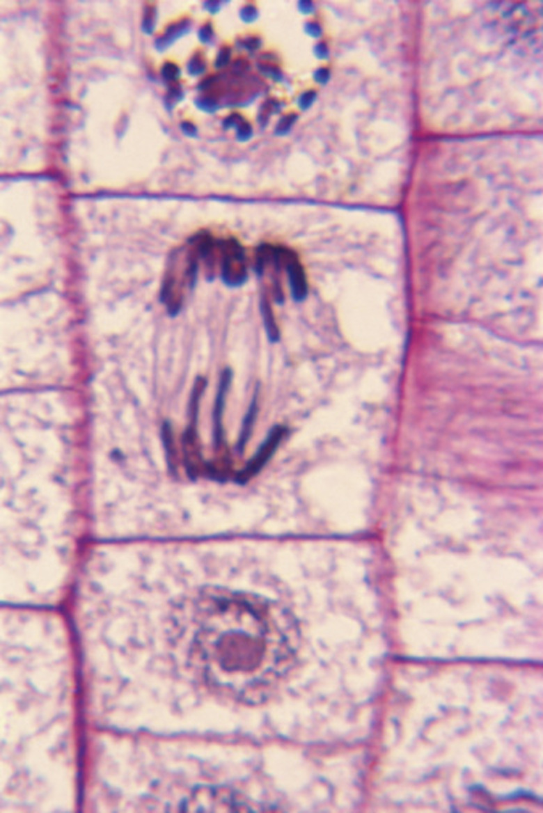
**Anaphase** usually only lasts a few moments and appears dramatic. This is the phase of mitosis during which the sister chromatids separate completely and move to opposite sides of the cell.
If you view early anaphase using a microscope, you will see the chromosomes clearly separating into two groups. If you are looking at late anaphase, these groups of chromosomes will be on opposite sides of the cell.
You may even notice the very beginning of a new cell membrane forming down the center of the cell between the spindle fibers.
Telophase Under a Microscope
Telophase
Under a Microscope
During the last of the mitosis phases, **telophase**, the spindle fibers disappear and the cell membrane forms between the two sides of the cell. Eventually, the cell divides completely into two separate daughter cells via **cytokinesis**.
When you look at a cell in telophase under a microscope, you will see the DNA at either pole. It may still be in its condensed state or thinning out. The new nucleoli may be visible, and you will note a cell membrane (or cell wall) between the two daughter cells.
Cite This Article
MLA
Mayer, Melissa. "How To Identify Stages Of Mitosis Within A Cell Under A Microscope" sciencing.com, https://www.sciencing.com/identify-within-cell-under-microscope-8479409/. 29 April 2019.
APA
Mayer, Melissa. (2019, April 29). How To Identify Stages Of Mitosis Within A Cell Under A Microscope. sciencing.com. Retrieved from https://www.sciencing.com/identify-within-cell-under-microscope-8479409/
Chicago
Mayer, Melissa. How To Identify Stages Of Mitosis Within A Cell Under A Microscope last modified August 30, 2022. https://www.sciencing.com/identify-within-cell-under-microscope-8479409/
